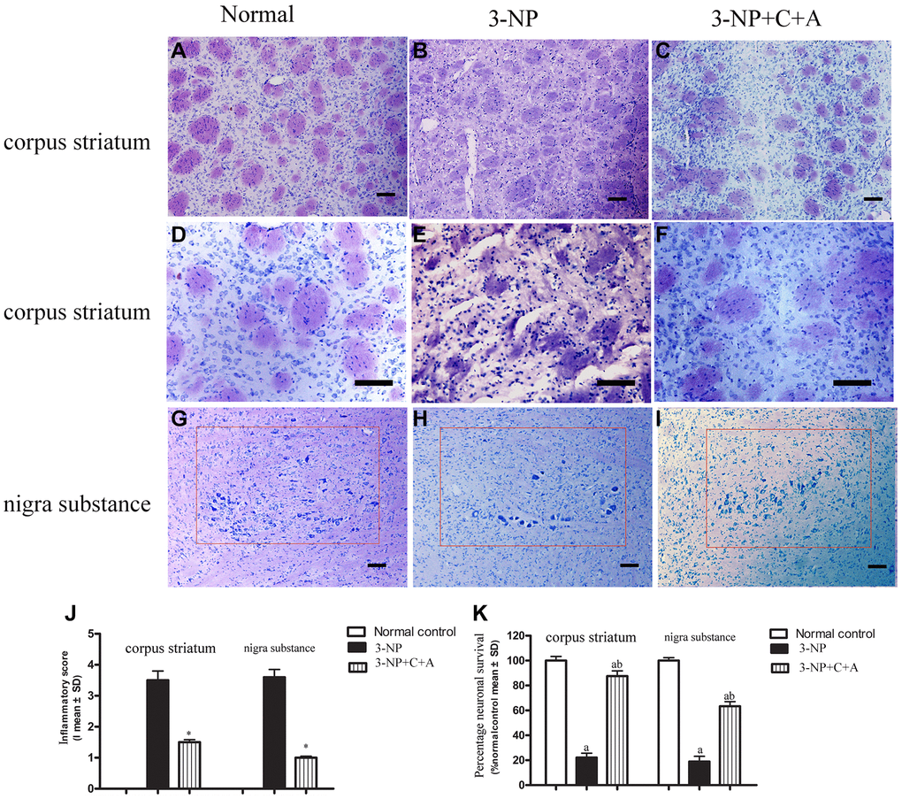
Figure 4.(A–F) HE staining showed inflammatory cells in the corpus striatum of each group at low magnification (×100, A–C) and higher magnification (×200, D–F); and (G–I) images of the surviving neurons within the nigra substance region (red box) for the control, 3-NP, and 3-NP+C16+Ang-1 groups. (J–K) Semi-quantitative profiles of the (J) inflammation scores (0, no inflammation; 1, cellular infiltrates only detected around meninges and blood vessels; 2, mild infiltrates detected in parenchymal tissues (1–10 inflammatory cells per slide); 3, moderate infiltrates observed in parenchymal tissues (11–100 inflammatory cells per slide); and 4, severe infiltrates in parenchymal tissues (>100 inflammatory cells per slide)) as well as the (K) neurons in each group visualized using a Nikon TE-300 microscope (Nikon, Japan) at ×200 magnification. The percentage (%) of neuronal survival was calculated with respect to the % of the sham control. Three randomly selected fields-of-view per section were selected for counting. The entire substantia nigra region comprising of surviving cells that were included in the count is also shown (within red box). Scale bar = 100 μm. aP < 0.05 versus control; bP < 0.05 versus 3-NP-treated mice. 3-NP, 3-NP; 3-NP+C+A, 3-NP+C16+Ang-1.