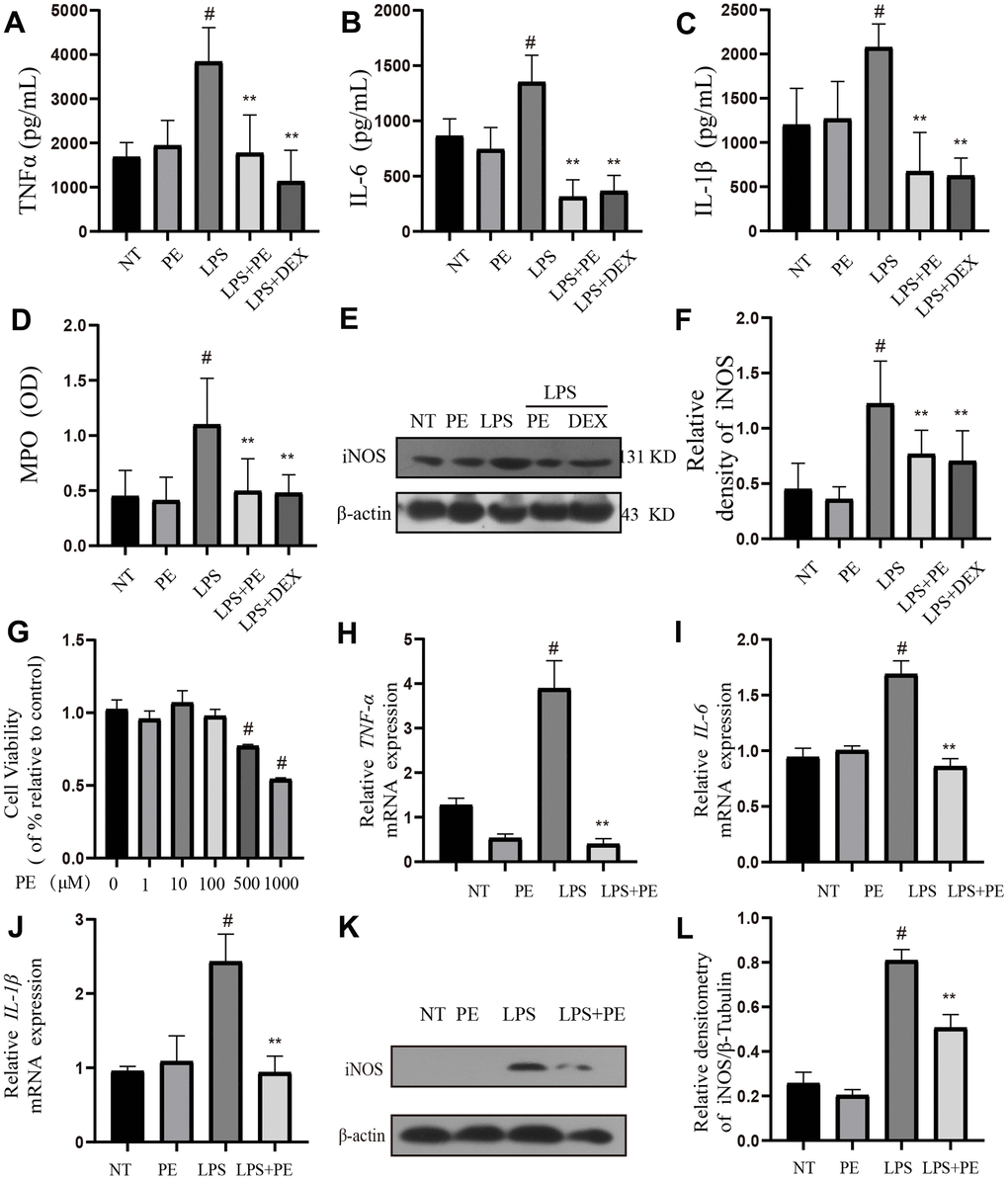
Figure 2.Effect of PE on inflammation level in mastitis model in vivo and in vitro. PE (10 mg/kg) was given orally for 7 days before the establishment of mastitis model. The fourth pair of milk ducts in mice were injected with 50 μL of 0.2 μg/μL LPS for 24 h. The mice were killed by dislocation and fixed on the operating platform. The hair was disinfected and fixed by spraying 70% alcohol. The midline of abdomen was cut to expose the breast tissue. Finally, the mammary was collected. mMECs was pretreatmented with 10 μM PE for 1 h, and then LPS stimulated the cells for 12 h. The inflammatory mediator’s levels of mice mammary gland and mouse mammary epithelial cells (mMECs) were measured by ELISA, MPO activity assay, western blot or qRT-PCR. (A–C) TNF-α, IL-6 and IL-1β protein content in mammary gland; (D) MPO content in mammary gland. (E, F) Western blot assay of iNOS protein in mammary tissue and in mMECs. (G) The activity of mMECs. (H–J) TNF-α, IL-6, IL-1β gene expression level in mMECs. (K, L) Western blot assay of iNOS protein in mMECs. Values are presented as means ± SEM, three independent repeated experiments were performed. #p<0.01 vs. NT group; **p < 0.01 vs. LPS group.