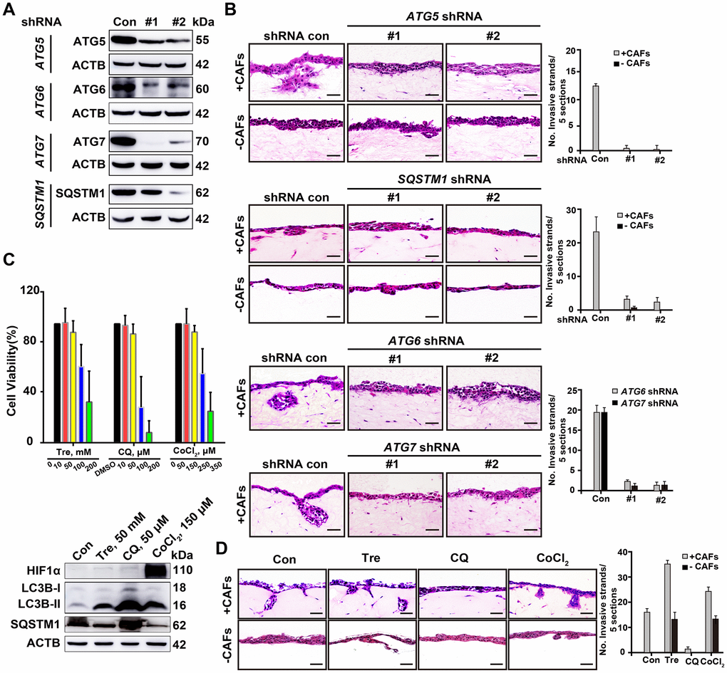
Figure 4.Deficient autophagy blocks cell invasion into organotypic gels. (A) The knockdown efficiency of shRNAs for autophagy-related proteins, including ATG5, ATG6, ATG7 and SQSTM1, was assessed by Western blotting. (B) Representative H&E-stained sections of ATG5-, ATG6-, ATG7- and SQSTM1-depleted A549 cells in the 3D organotypic coculture invasion system with or without human CAFs. The experiment was performed in triplicate. Scale bars, 50 μm. (C) The viability of A549 cells was evaluated by a CCK8 assay after 72 h of treatment with Tre, CQ, DMSO (vehicle control), or CoCl2 at the indicated concentration (upper panel). Error bars, means ± SD of a representative set of triplicate experiments. Immunoblots of HIF1α, LC3B, and SQSTM1 with the indicated treatment for 10 h, ACTB used as a loading control (lower panel). (D) The invasion capability of A549 cells was evaluated in the 3D organotypic coculture invasion system on gels with or without CAFs under treatment with DMSO, Tre (50 mM), CQ (50 μM), or CoCl2 (150 μM) for 10 days. The experiment was performed in triplicate. Scale bars, 50 μm. The number of invasive strands was quantified in 5 H&E-stained sections of each organotypic coculture gels. Error bars, means ± SD of a representative set of triplicate experiments.