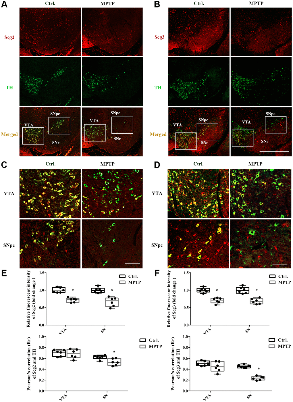
Figure 2.Double immunofluorescence staining of Scg2 or Scg3 with TH-positive DA neurons. (A–B) Co-staining of Scg2 or Scg3-positive secretory granules (Red) with TH-positive DA neurons (Green) at low magnification (20×), Scale bar, 50 mm. (C–D) Co-staining of Scg2 or Scg3-positive secretory granules (Red) with TH-positive DA neurons (Green) in the ventral tegmental area (VTA) and substantia nigra par compacta (SNpc) at high magnification (40×), Scale bar, 50 mm. (E–F) Scg2 or Scg3 protein expressions in substantia nigra of control and MPTP-induced PD mice were quantified by mean fluorescence. And the quantification of colocalization between Scg2 or Scg3 and TH was determined by Pearson’s correlation coefficient using the Image Pro Plus software. Two-tailed unpaired Student t-tests were performed between the control and treated groups. *Statistically significant with P < 0.05; Error bars are SD; N = 6.