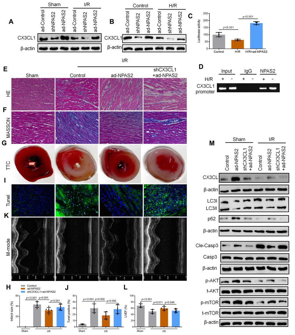
Figure 6.NPAS2 transcriptionally promoted CX3CL1 expression in vivo. (A) The protein level of CX3CL1 (100kDa) in rat myocardial tissue was determined by Western Blot. (B) The protein level of CX3CL1 (100kDa) in H9c2 cells was determined by Western Blot. (C) H9c2 cells were transfected with CX3CL1 promoter and the luciferase activity was determined after 24 hours. (D) Amplification of the CX3CL1 promoter sequence was performed BY ChIP assay in H9c2 cells. (E, F) Typical images of H&E and Masson staining of myocardial tissue segments. (G, H) Typical images of TTC of myocardial tissue segments. The infarct size was measured and calculated as a percentage of the total area. (I, J) Typical images of Tunnel of myocardial tissue segments. The relative percentages of apoptotic cells were calculated. (K, L) Typical echocardiographic images of M-mode and LVEF. (M) The protein level of CX3CL1 (100kDa), LC3B (14 and 16kDa), p62 (62kDa), Cleaved-Caspase-3 (17kDa), Caspase-3 (17kDa), p-AKT (60kDa), t-AKT (60kDa), p-mTOR (289kDa) and t-mTOR (289kDa) in rat myocardial tissue was determined by Western Blot. Data are expressed as mean ± SEM (n = 6).