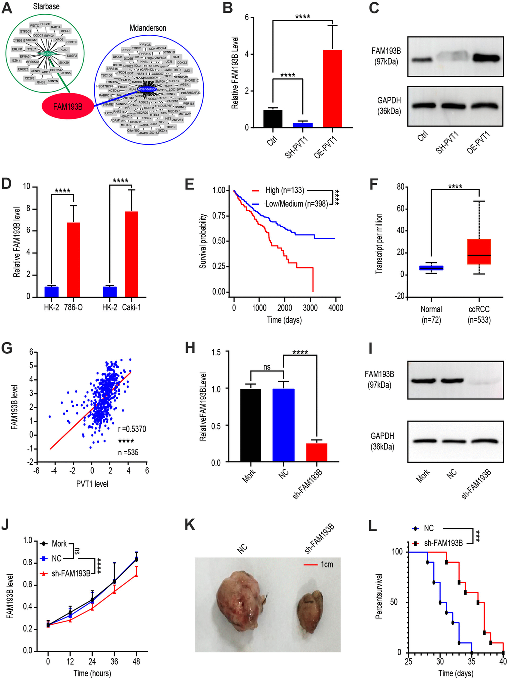
Figure 2.Effect of PVT1 target gene, FAM193B, on ccRCC cell proliferation in vitro and in vivo. (A) Potential target gene of PVT1 was calculated and overlapped according to Starbase and MD Anderson databases. (B) FAM193B levels were determined by qRT-PCR after treatment with lentivirus vector, SH-PVT1 lentivirus or OE-PVT1 lentivirus for 24 hours. (C) Protein levels of FAM193B were determined by western blot after treatment with lentivirus vector, SH-PVT1 Lentivirus, or OE-PVT1 Lentivirus for 48 hours. (D) Relative FAM193B levels in 786-O and Caki-1 cells compared to HK-2 cells. (E) ccRCC patients were divided into high FAM193B (n=398) and low FAM193B group (n=133) and survival times were recorded according to TCGA database. (F) Transcript levels of FAM193B in normal renal tissues (n=72) and ccRCC patients (n=533) according to TCGA database. (G) Interrelationship of PVT1 and FAM193B in ccRCC tissues (n=535) according to TCGA database. (H) FAM193B levels were determined by qRT-PCR after treatment with Mork, Lentivirus vector or SH-FAM193B Lentivirus for 24 hours. (I) Protein levels of FAM193B were determined by western blot after treatment with Mork, Lentivirus vector, or SH-FAM193B lentivirus for 48 hours. (J) Cell viability was determined by CCK-8 assay after treatment with Mork, Lentivirus vector, or SH-FAM193B lentivirus and recorded every 12 hours. (K) Tumor shape and size of ccRCC xenograft models harboring tumors generated by cells treated with Mork, Lentivirus vector, or SH-FAM193B lentivirus for 30 days. (L) Survival time of ccRCC xenograft models harboring tumors generated by cells treated with Mork. Mean ± SEM, ***P < 0.005, ****P < 0.001, NS: no significance.