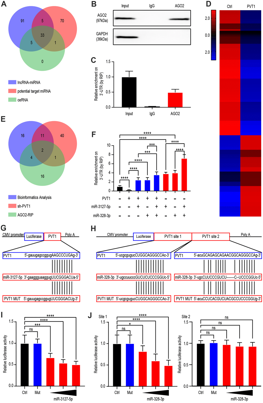
Figure 3.PVT1 regulated miR-3127-5p and miR-328-3p by serving as a sponge gene. (A) Venn diagram showing the over-lapping of potential target miRNAs of PVT1 according to Starbase database. (B) Co-IP and western blot assays indicating the interaction between AGO2 and HuR in AGS cells. (C) RIP and qRT-PCR assays revealing the endogenous binding of AGO2 to the 3’-UTR of target genes in Caki-1 cells. (D) Heatmaps showing 142 differentially expressed genes (fold change > 2.0, P < 0.05) in Caki-1 cells stably transfected with lentivirus vector or SH-PVT1 lentivirus for 24 hours. (E) Venn diagram showing the over-lapping of RNA-seq results from Caki-1 cells stably transfected with sh-PVT1 lentivirus, RIP assays, and potential target miRNAs from Figure 2A. (F) RIP and qRT-PCR assays revealing the endogenous binding 3’-UTR of target genes in Caki-1 cells transfected with Mock, PVT1, or miRNA mimics (100 nmol/L). (G) Schematic view of miR-3127-5p putative targeting site in the WT and MUT 3’UTR of PVT1. (H) Schematic view of miR-328-3p putative target site in the WT and MUT 3’UTR of PVT1. (I) Luciferase activity assay in 293 T cells transfected with luciferase reporter plasmids harboring PVT1 3’UTR (WT or MUT) and control miRNA or miR-3127-5p. (J) Luciferase activity assay in 293 T cells transfected with luciferase reporter plasmids harboring PVT1 3’UTR (WT or MUT) and control miRNA or miR-328-3p. Mean ± SEM, ***P < 0.005, ****P < 0.001.