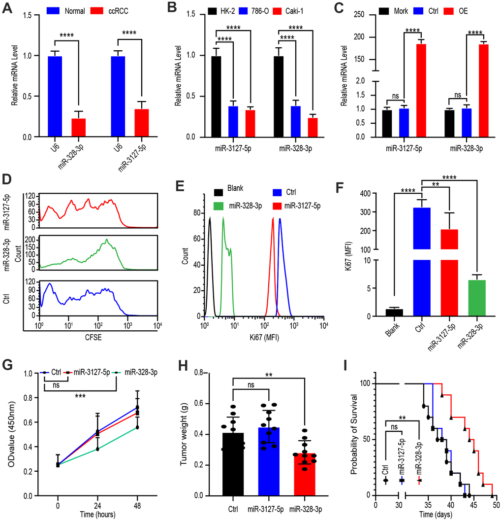
Figure 4.PVT1 promotes ccRCC proliferation mainly through miR-328-3p. (A) qRT-PCR assays of miR-328-3p and miR-3127-5p in ccRCC tissues and adjacent normal renal tissues (n=45). (B) Expression levels of miR-328-3p and miR-3127-5p in ccRCC cell lines, 786-O, and Caki-1 compared with those in the normal renal cell line, HK-2, were determined by qRT-PCR. (C) Caki-1 cells treated with Mork, Lentivirus vector, OE-miR-328-3p lentivirus, or OE-miR-3127-5p lentivirus for 24 hours; levels of miR-328-3p and miR-3127-5p were determined by qRT-PCR. (D) Fluorescence attenuation of CFSE-labeled Caki-1 cells after treatment with lentivirus vector, OE-miR-328-3p lentivirus, or OE-miR-3127-5p lentivirus for 48 hours. (E, F) Flow cytometry assays to determine MFI of Ki67 in Caki-1 cells treated with empty lentivirus vector, OE-miR-328-3p lentivirus, or OE-miR-3127-5p lentivirus for 48 hours. (G) Cell viability was determined by CCK-8 assays after treatment with lentivirus vector, OE-miR-328-3p lentivirus, or OE-miR-3127-5p lentivirus; data were recorded every 24 hours. (H) Tumor weight of ccRCC xenograft models harboring tumors generated by cells treated with lentivirus vector OE-miR-328-3p lentivirus or OE-miR-3127-5p lentivirus for 30 days. (I) Survival time of ccRCC xenograft models. Mean ± SEM, *P < 0.05, ** P < 0.01, ****P < 0.001, NS: no significance.