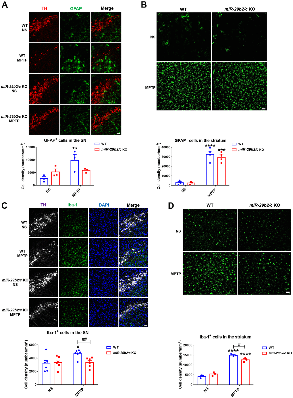
Figure 4.The analysis of glial activation in the nigrostriatal pathway at 3 days after MPTP administration. (A) Immunofluorescence staining for TH (red) and GFAP (green) in the SNpc of WT and miR-29b2/c KO mice. Scale bar: 0.1 mm. n =3-6. (B) Immunofluorescence staining for GFAP (green) in the striatum of WT and miR-29b2/c KO mice. Scale bar: 0.02mm. n =3-4. (C) Immunofluorescence staining for TH (white) and Iba-1 (green) in the SNpc of WT and miR-29b2/c KO mice. Nuclei were counterstained with DAPI (blue). Scale bar: 0.1 mm. n=6. (D) Immunofluorescence staining for Iba-1(green) in the striatum of WT and miR-29b2/c KO mice. Scale bar: 0.02mm. n=3-4. The counting of GFAP positive cells and Iba-1 positive cells in the SNpc and the striatum is shown in the lower panels. The differences were analyzed by two-way ANOVA followed by LSD multiple comparison tests. *p<0.05, **p<0.01, ***p<0.001 and ****p<0.0001, vs normal saline control. #p<0.05, vs WT group.