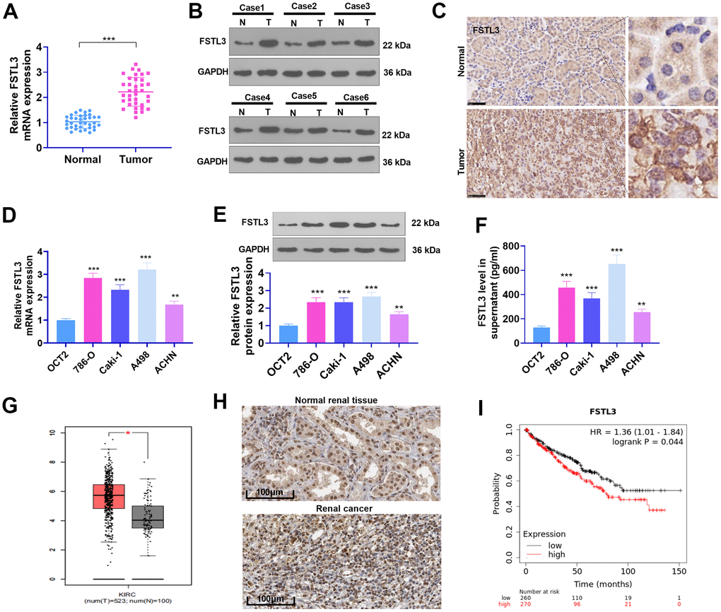
Figure 1.The profile of FSTL3 in RCC. (A) qRT-PCR determined FSTL3’s expression in RCC tissues as well as adjacent normal tissues, ***P<0.001 (vs. Normal group); (B) Western blot was taken for FSTL3 profile detection in RCC tissues and adjacent normal tissues. (C) IHC ascertained FSTL3’s expression in RCC tissues and adjacent normal tissues, Bar=50 μm; (D, E) qRT-PCR (D) and Western Blot (E) examined FSTL3 in the OCT2, A498, 786-O, Caki-1, and ACHN cell lines, **P<0.01, ***P<0.001 (vs. the OCT2 group). N=5. (F) ELISA measured FSTL3 in the OCT2, A498, 786-O, Caki-1, and ACHN culture mediums, **P<0.01, ***P<0.001 (vs. the OCT2 group). (G) FSTL3 expression in RCC was figured out through the GEPIA database (http://gepia.cancer-pku.cn/). (H) FSTL3’s expression in RCC tissues or normal renal tissues was analyzed via Human Protein Atlas (https://www.proteinatlas.org/). (I) The Kaplan-Meier Plotter database (http://kmplot.com/analysis/index.php?p) was consulted to confirm FSTL3 expression in the survival rate of KIRC patients.