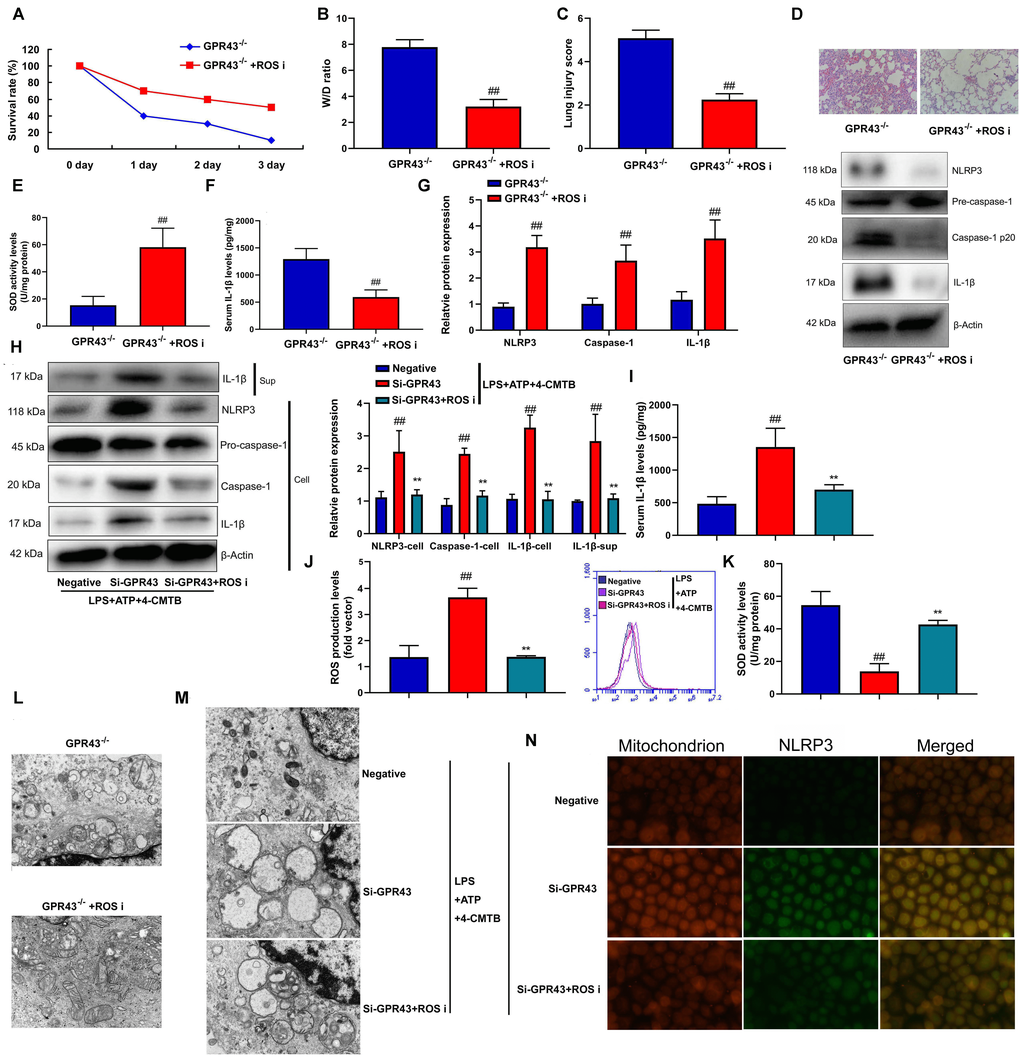
Figure 4.The inhibition of GPR43 activate NLRP3 inflammasome by ROS production-induced mitochondrial fission. Survival rate (A) in GRP43-/- mice with CLP and ROS I for 72 h; W/D rate (B), lung injury score (C), lung tissue using HE staining (D), SOD activity level (E), serum IL-1β levels (F), NLRP3/caspase-1/ IL-1β protein expressions (G) in GRP43-/- mice with CLP and ROS I for 24 h; NLRP3, Caspase-1 and IL-1β protein expressions in cells and IL-1β protein expression in supernatant (H), IL-1β levels (I), ROS production level (J), and SOD activity levels (K) in macrophage by down-regulation of GPR43 and LPS+ATP+GPR43 agonist for 24 h; Representative electron microscopy images (L) in macrophage of GRP43-/- mice with CLP for 24 h; Representative electron microscopy images (M) in macrophage by down-regulation of GPR43 and LPS+ATP+GPR43 agonist for 24 h; Confocal showed the accumulation of ROS production within mitochondria (N) in macrophage by down-regulation of GPR43 and LPS+ATP+GPR43 agonist for 24 h. GPR43-/-, GPR43-/- mice with CLP; GPR43-/-+ROS i, GPR43-/- mice of CLP with ROS inhibitor; Negative, negative control; Si-GPR43, down-regulation of GPR43; LPS+ATP+4-CMTB, macrophage by treated with LPS+ATP+4-CMTB. ##p<0.01 compared with GPR43-/- mice with CLP or GPR43-/- mice with CLP; **p<0.01 compared with down-regulation of GPR43.