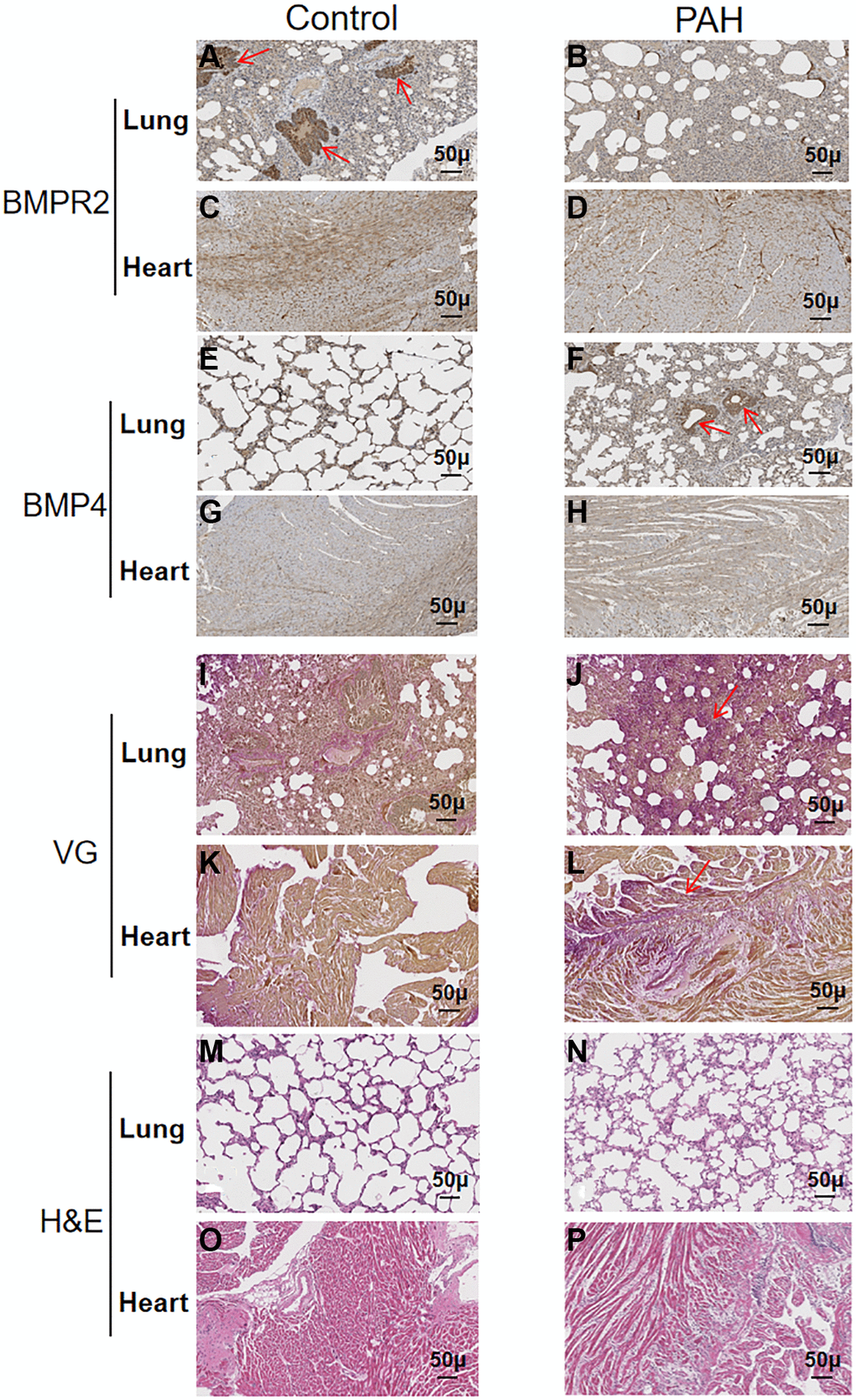
Figure 5.Reduced BMPR2 expression and higher fibrosis were observed in a pulmonary artery hypertension (PAH) mouse model. Abdominal shunt was used to create a pulmonary artery hypertension (PAH) mouse model. The lung and heart sections of 4 PAH mice were compared with those of 4 control mice. Scale bars, 50 μm. (A–D) BMPR2 expression was reduced on the lung section of the pulmonary hypertension model mice compared with that of the control mice based on immunohistochemistry staining. The brown stained areas indicated by the arrows in Figure 5A showed high BMPR2 protein levels in the lung section of the control mice. This reduction of BMPR2 was not observed in the heart sections comparing PAH mice with the control mice. (E–H) Higher BMP4 expression was observed on the lung section but not on the heart section of the PAH mice compared with the control mice by immunohistochemistry. The brown stained areas indicated by the arrows in Figure 5F showed high BMP4 protein levels on the lung section of the PAH mice. (I–L) Fibrosis was observed in the PAH mice. Van Gieson staining was carried out for the lung and heart section of the PAH and control mice. The area indicated by the arrow in Figure 5J and Figure 5L showed fibrosis on the lung and heart section of the PAH mice, respectively. (M–P) hematoxylin-eosin staining was carried out for the lung and heart section of the PAH and control mice.