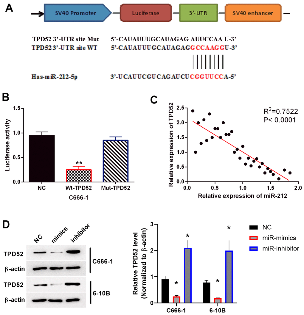
Figure 4.TPD52 was a direct target of miR-212. NC represented C666-1 cells with empty sequence. (A) Predicted binding site between TPD52 and miR-212. (B) Luciferase reporter assay with WT-TPD52 and Mut-TPD52. N=3. (C) miR-212 was negatively correlated with TPD52 in NPC tissues. N=33. (D) miR-212 mimics or inhibitor regulated TPD52 expression. The western blots were the representative images of 3 experiments. Data were quantified and normalized to the intensity of β-actin. **p <0.01.