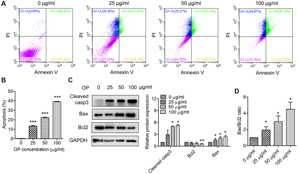
Figure 2.OPE enhances apoptosis in A20 cells. (A, B) Flow cytometry analysis of apoptosis of A20 cells after treatment with OPE (0, 25, 50, 100 μg/mL) for 48 h. (C) Western blot analysis of the expression levels of apoptosis-related proteins. A20 cells were treated with OPE (0, 25, 50, and 100 μg/mL) for 48 h, and Western blot was conducted with the indicated antibodies. (D) The alteration of the Bax/Bcl2 ratio in A20 cells following treatment with OPE. Data are presented as means ± SD of at least three independent experiments. (*p < 0.05; ***p < 0.001, compared to the untreated control).