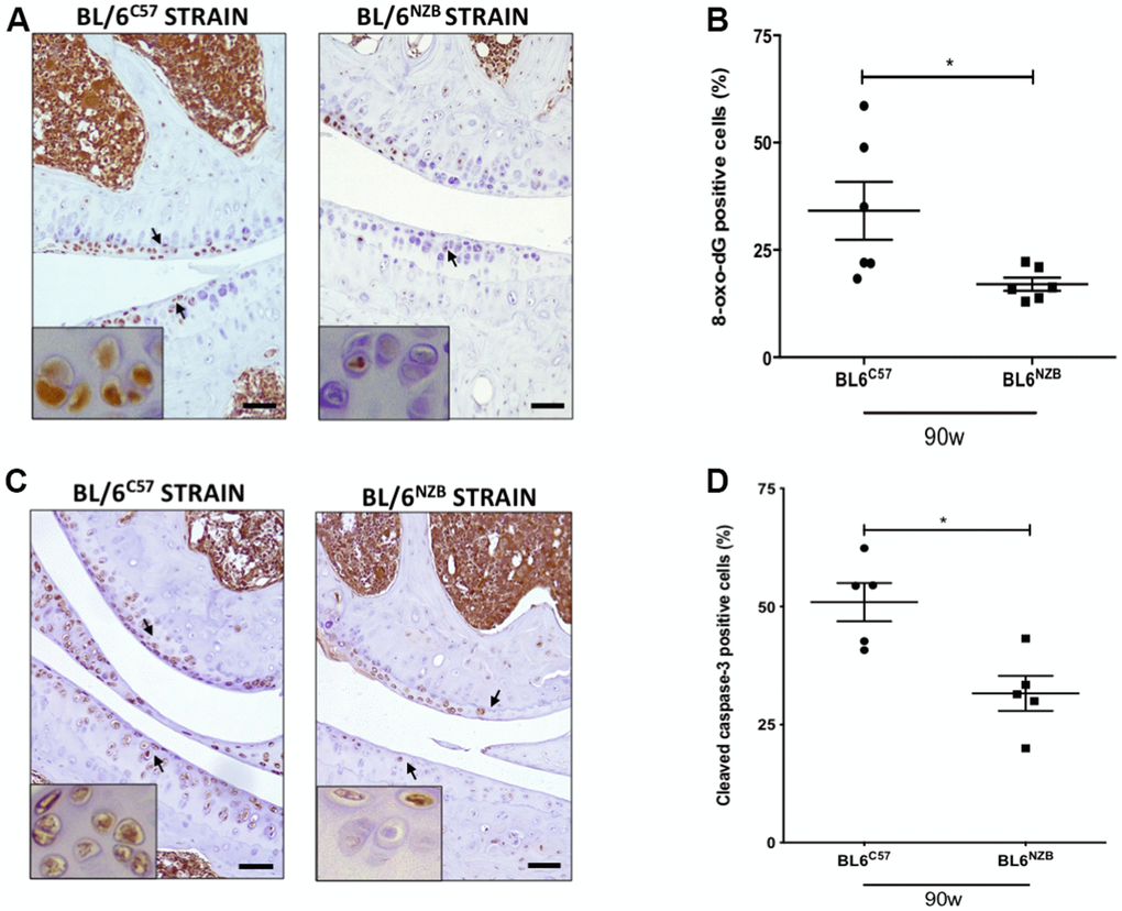
Figure 4.Reduction of 8-oxo-dG and cleaved caspase-3 expression in cartilage from conplastic (BL/6NZB) mice. (A) Representative images of medial compartment of knee joints from BL/6C57 and conplastic (BL/6NZB) mice at 90 weeks of age stained with 8-oxo-2′-deoxyguanosine (8-oxo-dG). (B) Quantitative analysis of 8-oxo-dG-positive cells of knee joints from BL/6C57 and conplastic (BL/6NZB) mice at 90 weeks. (C) Representative images of medial compartment of knee joints from BL/6C57 and conplastic (BL/6NZB) mice at 90 weeks of age stained with cleaved caspase-3. (D) Quantitative analysis of cleaved caspase-3-positive cells of knee joints from BL/6C57 and conplastic (BL/6NZB) mice at 90 weeks of age. Original magnification: 20×. Scale bar, 50 μm. Black arrow indicates positively stained chondrocyte. Chondrocyte magnification (40×) is shown in the bottom-left corner of the images. 8-oxo-dG: Graphs represent means ± SEM; n=6 in BL/6C57 and n=6 in conplastic (BL/6NZB) mice at 90 weeks of age. Cleaved caspase-3: Graphs represent means ± SEM; n=5 in BL/6C57 and n=5 in conplastic (BL/6NZB) mice at 90 weeks of age; *p<0.05 by non-parametric Mann-Whitney test.