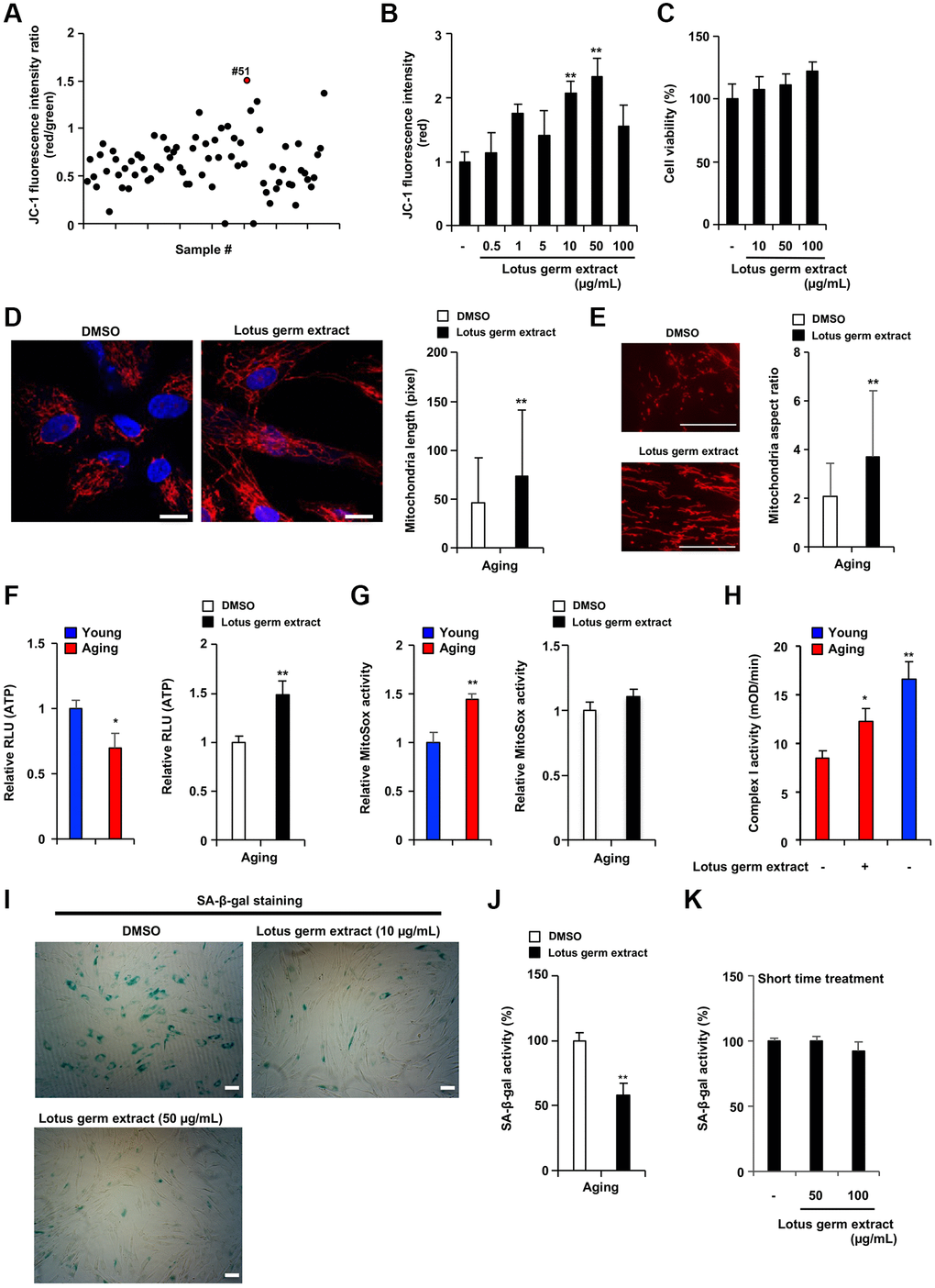
Figure 2.Lotus germ extract restored mitochondrial function and suppressed the aging phenotype. (A) Plant extract screening was performed in duplicate, and relative JC-1 activity was calculated using the procedure described in Figure 1C. (B and C) Effects of lotus germ extract on JC-1 activity and cell viability. Aging NB1RGB cells were treated with DMSO (−) or lotus germ extract at the indicated concentration for 24 h. (B) JC-1 activity was determined by the red fluorescence intensity, indicating activated mitochondria. (C) Cell viability was determined using an MTT assay. (D and E) Lotus germ extract induced mitochondrial morphological changes. Aging NB1RGB cells were treated with DMSO or 50 μg/mL lotus germ extract for 2 days, followed by treatment with MitoTracker Orange and fluorescence microscopy. Red represents mitochondrial MitoTracker staining, and blue represents nuclear DAPI staining (scale bar, 20 μm). (D) The length of the mitochondria within the cells was determined, and the data are presented as the mean ± SD (n = 30) (right panel). (E) The aspect ratio of the mitochondria within the cells was determined. The data are presented as the mean ± SD (DMSO: n = 83, lotus germ extract: n = 53) (right panel). (F and G) Lotus germ extract stimulated ATP but not ROS production. Young and aging cell were compared (left panel). Aging NB1RGB cells were treated with DMSO or 50 μg/mL lotus germ extract for 24 h (right panel). (F) ATP levels were determined using a CellTiter-Glo assay. (G) The ROS level was determined MitoSOX staining assay. (H) Mitochondrial complex I activity is stimulated by the lotus germ extract. Young NB1RGB cells treated with DMSO (−) or NB1RGB cells treated with DMSO (−) or 50 μg/mL of lotus germ extract (+) for 24 h were subsequently assessed for mitochondrial respiratory complex I activity. mOD, millioptical density. (I) Lotus germ extract decreased SA-β-gal-positive cells. Aging NB1RGB cells were treated with the indicated concentration of lotus germ extract for 3 days and stained to detect SA-β-gal-positive cells. (J and K) Lotus germ extract decreased SA-β-gal expression but did not directly suppress SA-β-gal activity. (J) Aging NB1RGB cells were treated with the indicated concentration of lotus germ extract for 3 days, and SA-β-gal activity was measured. (K) Aging NB1RGB cells were treated with DMSO (−) or the indicated concentration of the lotus germ extract for 2 h. Then, the SA-β-gal activity was measured. Data are presented as the mean ± SD of three simultaneously performed experiments (B–H, J and K). Each P value was calculated using two-way ANOVA; *P < 0.05, **P < 0.01.