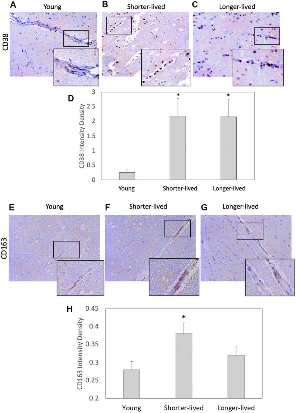
Figure 2.Expression of the inflammation markers CD38 and CD163 in PFC from young and elder animals. Paraffin-embedded PFC sections were stained with (A–C) antibody anti- CD38 (observed in brown color) and (E–G) anti- CD163, which were (D, H) digitally imaged for density intensity quantification in 8-bit binary masks using Image J (NIH). Rectangles indicate areas expanded for detail. (A) Representative CD38 image of young (4-7 yo) group, (B) representative CD38 image of shorter-lived elder animals. (C) Representative CD38 image of longer-lived elders. (D) CD38 staining density intensity measured in 8-bit digital whole section images and binary masks, using ImageJ. (E) Representative CD163 image of young (4-7 yo) group, (F) representative CD163 image of shorter-lived elder animals. (G) Representative CD163 image of longer-lived elders. (H) CD163 staining density intensity measured in 8-bit digital whole section images and binary masks, using ImageJ. All images are 40X magnification. N=4/group. *p<0.05 in multiple comparisons.