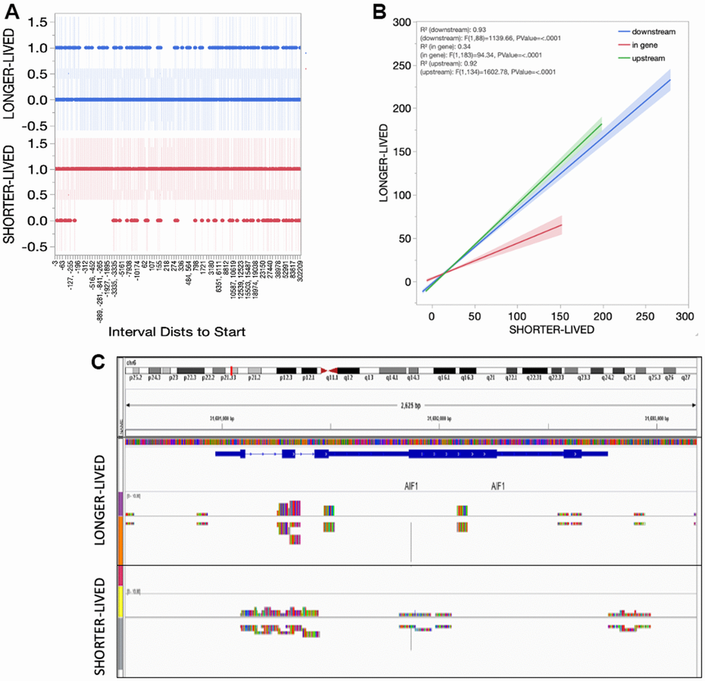
Figure 5.Sirt1 distribution in the PFC tissue from shorter- and longer- lived elder animals. (A) Interval distances from start where Sirt1 peaks are observed in chromatin preparations from PFC bulk tissue indicating presence (1) versus absence (0). (B) Interval positions of Sirt1 binding in PFC from shorter- and longer-lived animals. (C) Example of Sirt1 peak signal in the AIF1 gene, indicating a spread in shorter animals.