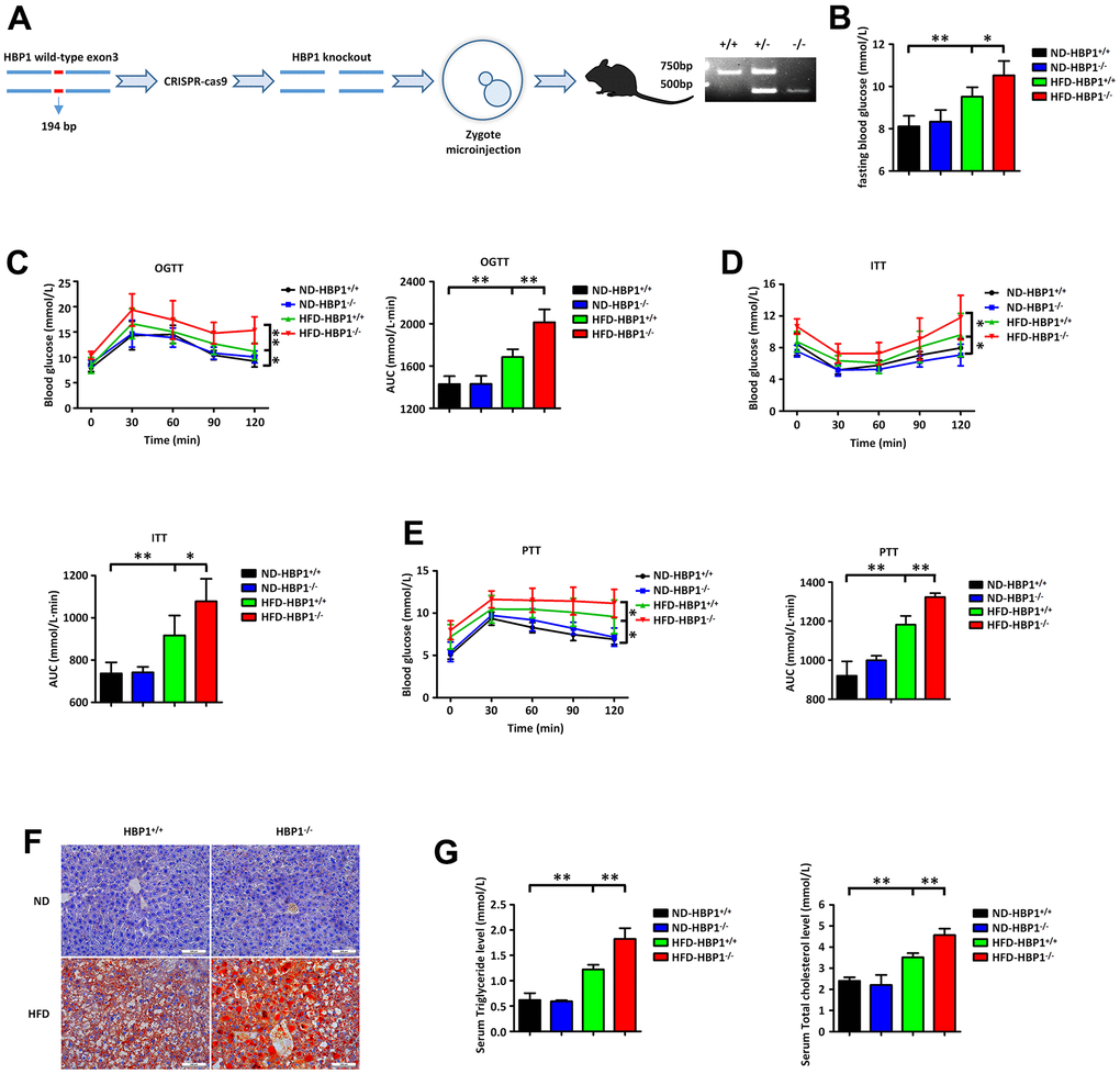
Figure 2.HBP1 knockout mice show a significant worsening of diabetes. (A) Schematic of the HBP1 knockout (HBP1−/−) mice and identification of genotypes. The genotypes were identified by PCR. The PCR product of wild type mice has only one band at the position of 611bp. The PCR product of heterozygous mice has one band at the position of 611bp and another one at the position of 417bp. The PCR product of HBP1−/− mice has only one band at the position of 417bp. (B) HBP1−/− mice fed by HFD has higher fasting blood glucose. Wild type C57BL/6J (HBP1+/+) mice and HBP1−/− mice of the same age were both divided into two groups randomly. One group was fed by ND as a control and the other was fed by HFD for 3 months to induce to diabetic mice. These four groups were fasted for 6 hours and drank water freely. After that, blood was collected from the tail vein of mice and its glucose content was measured by Roche glucometer. (C) HFD-HBP1−/− mice have more severe impaired glucose tolerance. All the four groups were fasted for 6 hours and drank water freely. After 6 hours, fasting blood glucose was recorded as 0 min point. Each mouse was given 5 mg/kg glucose by gavage and its glucose content in tail vein blood was measured by Roche glucometer every 30 minutes, up to 2 hours. (D) HFD-HBP1−/− mice are less sensitive to insulin. All the four groups were fasted for 4 hours and drank water freely. After 4 hours, fasting blood glucose was recorded as 0 min point. Each mouse was given 1 U/kg insulin by intraperitoneal injection and its glucose content in tail vein blood was measured by Roche glucometer every 30 minutes, up to 2 hours. (E) HFD-HBP1−/− mice have higher glucose excursion during pyruvate tolerance test. All the four groups were fasted for 16 hours and drank water freely. After 16 hours, fasting blood glucose was recorded as 0 min point. Each mouse was given 1 g/kg pyruvate by intraperitoneal injection and its glucose content in tail vein blood was measured by Roche glucometer every 30 minutes, up to 2 hours. (F) The livers of HFD-HBP1−/− mice have more severe steatosis. Representative photographs of liver samples were selected and analyzed by oil red O staining. Scale bar, 100 μm. (G) Serum triglyceride and total cholesterol contents in HFD-HBP1−/− mice are significantly increased. Serums were isolated from the inferior vena cava. AUC, Area Under Curve. Data were the mean ± SD by a one-way ANOVA. *, p<0.05. **, p<0.01.