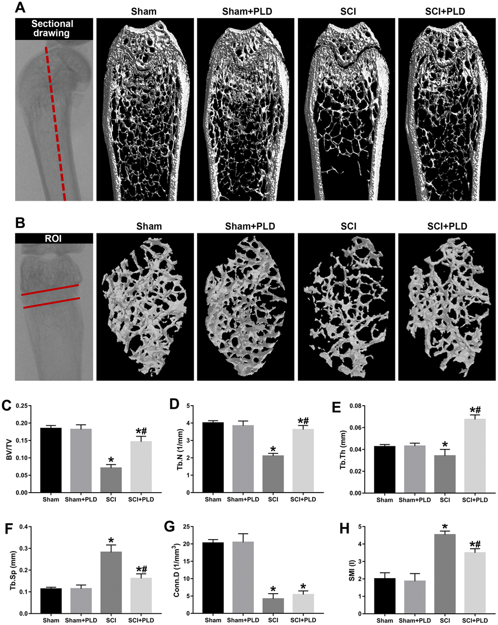
Figure 2.Effects of PLD on trabecular bone structure of the distal femur. (A) The schematic diagram of the coronal section of femur and representative 3D reconstructed coronal images of cancellous bone in the distal femur of each group. (B) The schematic diagram of the ROI of distal femur and the representative μCT 3D-images of cancellous bone within the ROI. Plots of the structural parameters: (C) BV/TV, (D) Tb.N, (E) Tb.Th, (F) Tb.Sp, (G) Conn.D, and (H) SMI. Data are expressed as mean ± S.D.; n=4 to 5 per group; *P < 0.05, vs. the Sham group; #P < 0.05, vs. the SCI group.