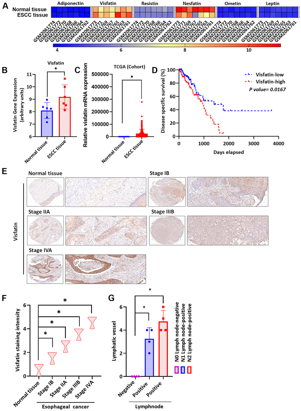
Figure 1.Clinicopathologic features of visfatin expression in human ESCC tissue samples. (A) The gene expression profiles of visfatin in ESCC tissue and normal tissue samples were analyzed in specimens from the GEO and TCGA databases. (B, C) Levels of visfatin were significantly increased in ESCC samples compared with normal tissue samples. (D) Kaplan-Meier analysis of overall survival according to visfatin expression in patients with esophageal cancer. (E, F) The human ESCC tissue array specimens were subjected to IHC evaluations with visfatin antibody, and levels of positive staining were quantified by IHC scoring (N = 4 per group). Scale bar: 100 μm. (G) Positive peritumoral lymphatic vessel density in patients with N0, N1, or N2 ESCC tissue array samples (N = 4 per group). *P < 0.05 compared with normal tissue samples or N0 lymph node negative tissue array samples.