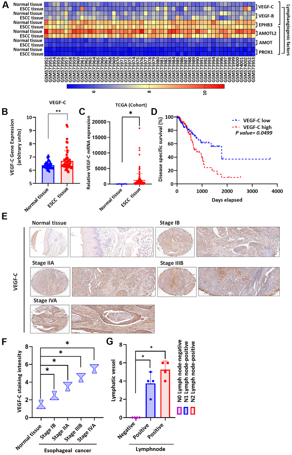
Figure 2.Clinicopathologic features of VEGF-C expression in human ESCC tissue. (A) The gene expression profiles of VEGF-C in ESCC tissue and normal tissue samples were analyzed in GEO and TCGA database records. (B, C) Levels of VEGF-C expression were significantly higher in ESCC samples compared with the normal tissue samples. (D) Kaplan-Meier analysis of overall survival according to VEGF-C expression in patients with esophageal cancer. (E, F) The human ESCC tissue array specimens were subjected to IHC evaluations with VEGF-C antibody, and the positive staining was quantified by IHC scoring (N = 4 per group). Scale bar: 100 μm. (G) Positive peritumoral lymphatic vessel density in patients with N0, N1, or N2 ESCC tissue array samples (N = 4 per group). *P < 0.05 compared with normal tissue samples or N0 lymph node negative tissue array samples.