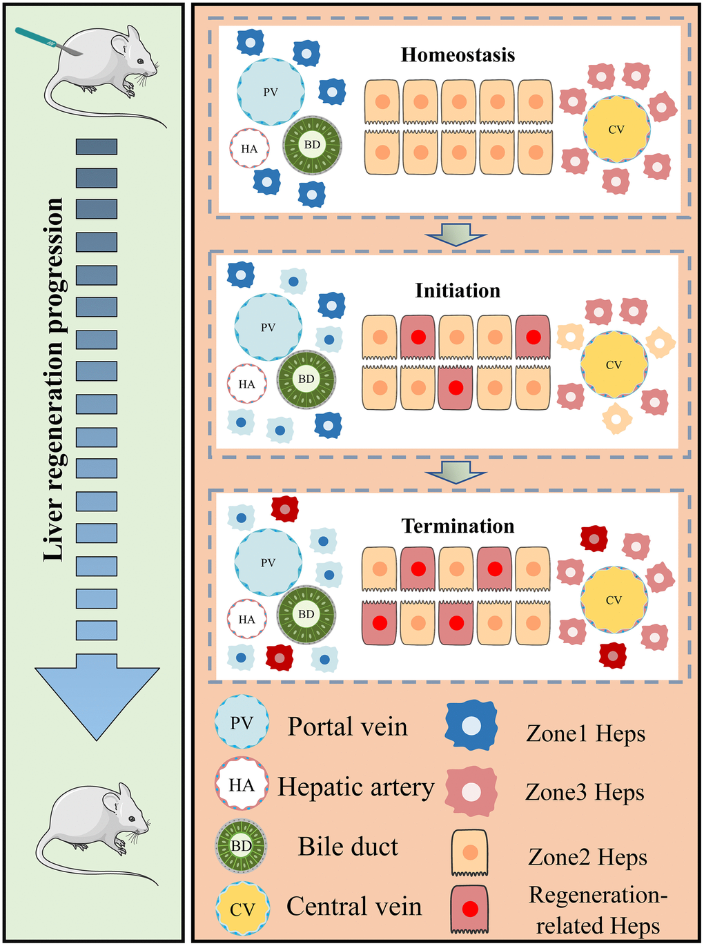
Figure 10.Regional characteristics of live regeneration-related hepatocytes during acute liver injury. The left panel of our study demonstrates the process of liver regeneration following PHx. The surgical resection of liver tissue triggers the rapid re-entry of remnant hepatocytes into the regeneration state. The right panel summarizes the zonal variation in regeneration-related hepatocytes during acute liver injury (AIL). Under homeostatic conditions, hepatocytes in different zones remain in a quiescent state. Portal vein (PV) and central vein (CV) hepatocytes are located at both ends of the hepatic lobules and exhibit distinct spatial gene expression patterns. However, during liver damage, newborn hepatocytes initiate in the midlobular regions before progressing towards the periportal and pericentral areas. At the same time, zonal differences in PV and CV hepatocyte gene expression decrease as hepatocytes respond to the regenerative challenge (hepatocytes with blue and yellow dim out). During the termination phase, newborn hepatocytes repopulate different regions, leading to the recovery of liver function.