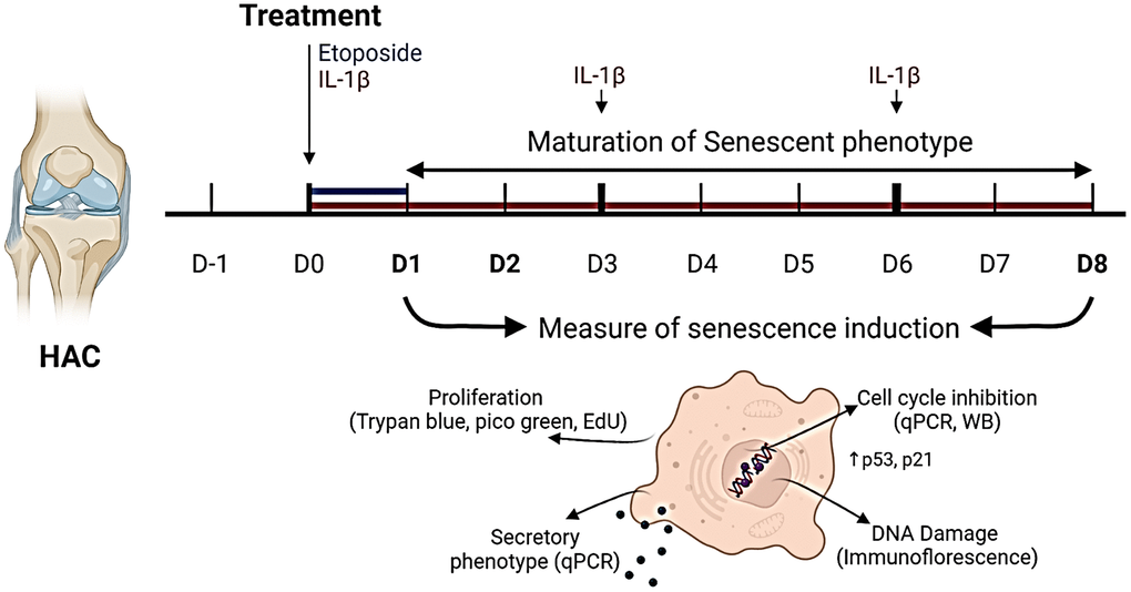
Figure 1.Experimental design. To investigate senescence in chondrocytes, primary human articular chondrocytes (HACs) were stimulated with etoposide for 24 h (blue) or with IL-1β for 8 days (red) with treatment renewal at days 3 and 6. Senescence features were assessed at days 1 and 8 in both conditions by qPCR, WB, and immunofluorescence. Figure created with https://www.biorender.com.
Figure 1 — Development of a DNA damage-induced senescence model in osteoarthritic chondrocytes | Aging