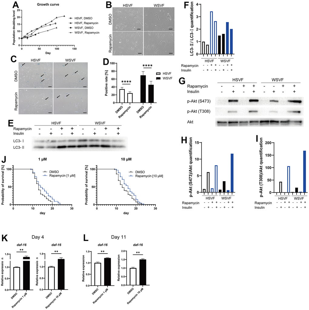
Figure 6.Rapamycin alleviates cellular senescence in SVF. (A) Growth curves of HSVF and WSVF treated with rapamycin. (B) Morphological changes of the SVFs treated with rapamycin. Scale bar, 100 μm. (C) Representative images of SA-β-gal staining of the SVFs treated with rapamycin. Black arrows indicate SA-β-gal-positive cells. Scale bar, 100 μm. (D) Quantification of SA-β-gal-positive cells. Data are presented as means ± S.E.M. from nine different microscopic views. For statistical analysis, student t-test was performed (****p < 0.0001). (E) Western blotting of the protein expression of LC3-I and LC3-II in HSVF and WSVF. (F) Quantification of (E). (G) Western blotting of p-Akt (S473), p-Akt (T308), and Akt in HSVF and WSVF treated with rapamycin. (H) Quantitative analysis of p-Akt (S473)/Akt. (I) Quantitative analysis of p-Akt (T308)/Akt. (J) Survival probability of WRN-knockout C. elegans (gk99) treated with 1 μM and 10 μM of rapamycin. For statistical analysis, log-rank (Mantel-Cox) test was performed; **p < 0.01 compared with DMSO in 1 μM of rapamycin, *p < 0.05 compared with DMSO in 10 μM of rapamycin. (K, L) Quantitative real-time polymerase chain reaction of the relative expression of daf-16 on days 4 and 11 in gk99 treated with 1 μM and 10 μM of rapamycin. Data are presented as means ± S.E.M. of three technical replicates. For statistical analysis, student t-test was performed (**p < 0.01). WS: Werner syndrome; SVF: stromal vascular fraction; HSVF: SVF derived from a healthy patient; WSVF: SVF derived from a patient with WS; SA-β-gal: senescence-associated β-galactosidase; mTORC1: mammalian target of rapamycin complex 1.