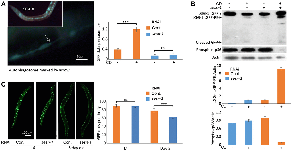
Figure 3.Autophagy activation by sesn-1 under CD correlates with reduced muscle degeneration. (A) Autophagosome accumulation in seam cells. Both control adIs2122 (DA2123 strain) and adIs2122; sesn-1(RNAi) nematodes, expressing a GFP-tagged LGG-1 fusion protein during L3, were exposed to axenic medium. Autophagosome counts per seam cell were analyzed under control conditions (n = 137 for adIs2122, n = 56 for adIs2122; sesn-1(RNAi)) and starvation conditions (n = 117 for adIs2122, n = 80 for adIs2122; sesn-1(RNAi)). “ns” and “***” indicate P-values > 0.05 and < 0.001, respectively. All bar graphs are presented as mean ± S.E.M. (B) Immunoblot and densitometric analyses showing relative levels of GFP::LGG-1, its phosphatidylethanolamine-conjugated form (LGG-1::GFP-PE), and the phosphorylated form of ribosomal protein S6 (phospho-rpS6) in adIs2122 (DA2123 strain) and adIs2122; sesn-1(RNAi) worms. All bar graphs represent blot intensity normalized to actin. (C) Whole-body images of nematodes expressing a myo-3p::GFP NLS-tagged fusion protein in body wall muscle nuclei in the ccIs4251 (PD4251 strain). Nuclear counts were performed at L4 (n = 24 for ccIs4251 and n = 23 for ccIs4251; sesn-1(RNAi)) and at 5 days of adulthood (n = 20 for both groups). “ns” and “***” indicate P-values > 0.05 and < 0.001, respectively. All bar graphs are presented as mean ± S.E.M.