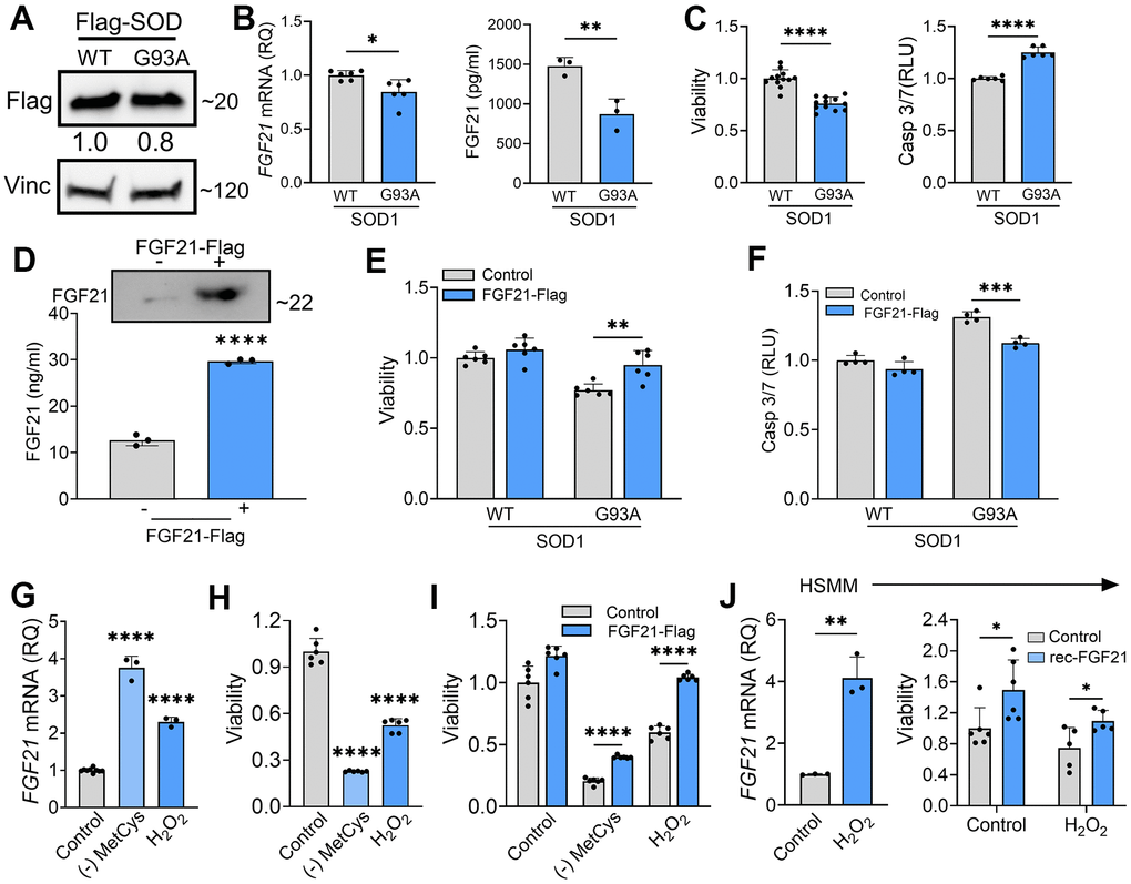
Figure 7.FGF21 mitigates cytotoxicity in C2C12 and human primary myoblasts induced by oxidative stress. (A) C2C12 myoblasts were transfected with FLAG-tagged WT and SOD1G93A expression plasmids and lysates were assessed by western blot using the antibodies indicated. Bands were quantitated by densitometry and a ratio to the loading control, vinculin, was calculated (shown between the two blots). (B) FGF21 mRNA levels were measured in the same lysates (left panel) and FGF21 protein in the conditioned media was quantified by ELISA (right panel). *P = 0.011, **P = 0.008; unpaired two-tailed t-test. Secretory FGF21 from the conditioned media was quantified using ELISA (right panel). (C) Viability of NSC-34 cells expressing FLAG-tagged WT-SOD1 or SOD1G93A was determined using the Vialight assay (left panel). Caspase activation was measured in the same cells and values were normalized to activity in WT SOD1-transfected cells which was set at 1 (right panel). ****P < 0.0001, unpaired two-tailed t-test. (D) FGF21 protein in the conditioned media of C2C12 cells transfected with a FLAG-tagged FGF21 was detected by western blot (upper panel) and by ELISA (graph). Estimated size of the band (kDa) is shown to the right of the blot. ****P < 0.0001; unpaired two-tailed t-test. (E, F) C2C12 myoblasts cells expressing either WT-SOD1 or SOD1G93A were transfected with FGF21-FLAG and assessed for viability and Caspase-3/7 activity as in (C). **P = 0.003, ***P = 0.0003; unpaired two-tailed t-test. (G) FGF21 mRNA levels were quantified in C2C12 cells exposed to MetCys-deprived media or treated with 100 mM H2O2 for 24 h. ****P < 0.0001; one-way ANOVA followed by Tukey’s multiple comparisons test. (H) Cell viability was assessed in C2C12 myoblasts exposed to stressors as described in (G). ****P < 0.0001; one-way ANOVA followed by Tukey’s multiple comparisons test. (I) C2C12 cells transfected with FGF21-FLAG (or empty vector) were subjected to stressors as described in (G) for 24h and then assayed for viability. ****P < 0.0001; unpaired two-tailed t-test. (J) FGF21 mRNA levels were quantified in human primary myoblast cells (HSMM) treated with 25 mM H2O2 for 24 h. **P = 0.0014; unpaired two-tailed t-test (left panel). HSMM cells treated with or without 25 mM H2O2 and recombinant FGF21 (100ng/ml) for 24h and then assayed for viability. *P < 0.05; unpaired two-tailed t-test (right panel). Data points represent biological replicates and bars are the mean ± SD.