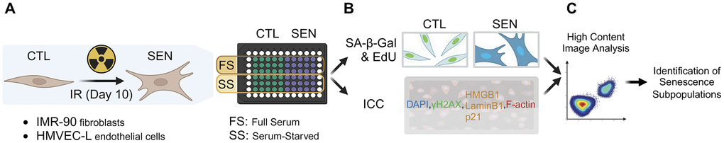
Figure 1.High-content imaging workflow. (A) Sample preparation. Human lung primary microvascular endothelial cells (HMVEC-L) and fibroblasts (IMR-90) were induced to senescence using ionizing radiation (IR). IR and mock-irradiated cells (CTL) were either cultured in full-serum medium the entire time (FS) or switched to low-serum medium for the last 3 days of culture to induce quiescence in CTL cells (SS). (B) Staining for senescence markers. Prepared samples were either co-stained for senescence-associated beta-galactosidase activity (SA-β-Gal) and proliferation via EdU incorporation (EdU); or for other senescence markers (γH2AX, LaminB1, HMGB1, p21) using immunocytochemistry (ICC). (C) High-content image analysis was performed to identify senescent subpopulations.