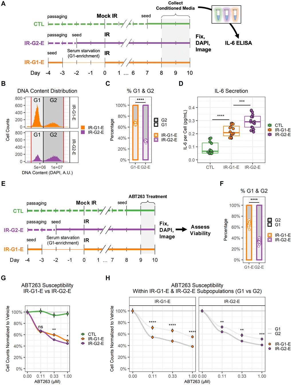
Figure 5.G1 and G2 senescent endothelial cells show different levels of IL-6 secretion and ABT263 susceptibility. (A) Workflow for the comparison of IL-6 secretion in G1 vs. G2 senescent endothelial cells. Three conditions were prepared: non-senescent mock-irradiated cells (CTL, green); ionizing radiation-induced senescent cells enriched in G2 (IR-G2-E, purple); ionizing radiation-induced senescent cells enriched in G1 (IR-G1-E, orange). Condition media (CM) were collected from the last 2 days of culture, after which cells were fixed, counterstained with DAPI, and imaged. IL-6 concentration was quantified by ELISA and normalized to cell counts. (B) DNA content distribution histogram, showing G1 (light grey) and G2 (dark grey) peaks in IR-G2-E and IR-G1-E senescent populations. The plot shows all IR-G2-E and IR-G1-E cells from a single representative experiment. (C) Quantification of (B), showing the sample percentages of senescent cells arrested in G1 and G2. Data shown is from 3 independent experiments; each data point is a sample (CTL n = 12; IR-G1-E and IR-G2-E n = 16). (D) IL-6 secretion across CTL, IR-G2-E, and IR-G1-E groups normalized to cell counts. Data shown are from 3 independent experiments; each data point is a sample (CTL n = 12; IR-G1-E and IR-G2-E n = 16). ***: p-value < 10-3; ***: p-value < 10-4; by one-way ANOVA followed by post-hoc Tukey’s test. (E) Workflow for the comparison of ABT263 susceptibility in G1 vs. G2 senescent endothelial cells. CTL, IR-G2-E, and IR-G1-E were prepared as described in (A), but cells were treated with ABT263 for the last 24 h of culture. After treatment, cells were fixed, counterstained with DAPI, and imaged. (F) Percentages of senescent cells per well arrested in G1 and G2. Data shown are from 3 independent experiments; each data point is a well (n = 30). (G) Cell viability comparison after ABT263 treatment between IR-G2-E and IR-G1-E senescent populations, measured by cell counts normalized to vehicle (0.00 μM ABT263). Data shown are mean ± SEM for each ABT263 concentration from 3 independent experiments. Viability was compared between IR-G2-E and IR-G1-E populations across all ABT263 concentrations (n = 30). ns: p-value > 0.05; *: p-value < 0.05; **: p-value < 0.01; by non-parametric Mann-Whitney test corrected for multiple comparisons by FDR method. (H) Cell viability comparison of G1 and G2 subpopulations within IR-G2-E and IR-G1-E populations from (G). Data shown are mean ± SEM of G1 (light grey) and G2 (dark grey) subpopulations for each ABT263 concentration from 3 independent experiments. Viability was compared between G1 and G2 cells across all ABT263 concentrations (n = 30). **: p-value < 10-2; ***: p-value < 10-3; ****: p-value < 10-4; by non-parametric Mann-Whitney test corrected for multiple comparisons by FDR method.