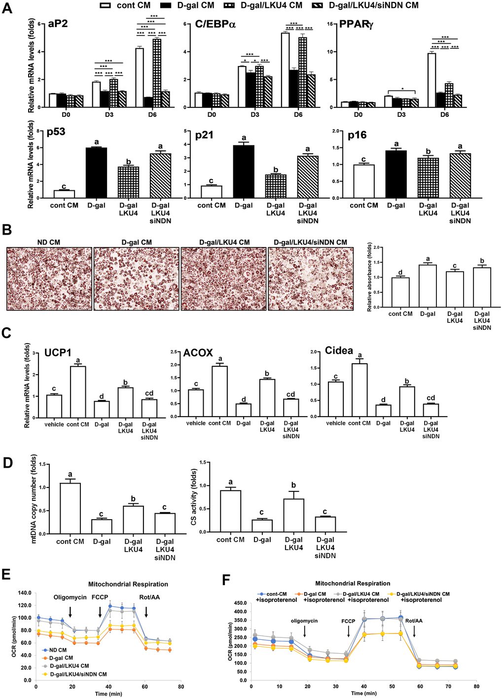
Figure 6.NDN knockdown facilitates age-related changes in cell functions in iWAT. (A–E) SVF and 3T3-L1 cells were differentiated in the absence or presence of CMs from iWAT explants. NDN knockdown was performed ex vivo in the iWAT explants from D-gal/LKU4 mice using NDN-specific siRNA. (A) RT-qPCR analysis of adipogenic genes during adipocyte differentiation. (B) Lipid accumulation was determined at day 6 by ORO staining. Scale bars, 200 μm. (C) On day 6, primary adipocytes differentiated from SVF cells were treated with 100 μM isoproterenol for 48 h. RT-qPCR was performed to analyze the expression of browning marker genes. (D) Mitochondria function was determined by measuring mtDNA copy number and CS activity in day 6 primary adipocytes. (E) OCR analysis of day 6 primary adipocytes and (F) OCR analysis of day 8 primary adipocytes treated with 100 μM isoproterenol for 48 h, were performed in the presence of oligomycin, FCCP, and rotenone/antimycin A using a Seahorse XFe analyzer at the indicated timepoints. All data are expressed as the mean ± S.E.M. * p < 0.05, *** p < 0.01. The lowercase letters above the graphs indicate statistical significance at p < 0.05.