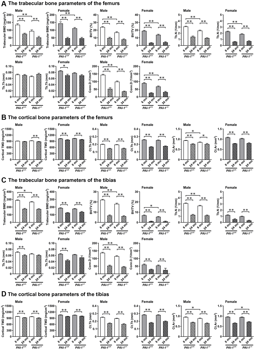
Figure 3.Aging-related changes in the microarchitecture of long bones in male and female mice. Trabecular and cortical bone parameters in the femurs (A, B) and tibias (C, D) of 6- and 24-month-old mice with PAI-1 gene deficiency (PAI-1−/−) and their wild-type counterparts (PAI-1+/+) are shown (n = 5−10 in each group). The distal metaphyseal regions of the femur and proximal metaphyseal regions of the tibia were analyzed using μCT. Trabecular bone parameters: trabecular bone mineral density (BMD), ratio of the segmented trabecular bone volume to the total tissue volume of the region of interest (BV/TV), trabecular number (Tb.N), trabecular thickness (Tb.Th), and connectivity density (Conn.D). Cortical bone parameters: cortical tissue mineral density (TMD), cortical thickness (Ct.Th), and cortical bone area (Ct.Ar). Data are presented as mean ± standard error of the mean. **P < 0.01 and *P < 0.05.