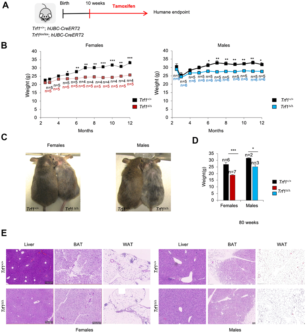
Figure 1.Depletion of Trf1 in mice induces weight loss. (A) Experimental plan: Trf1+/+ or Trf1lox/lox; hUBC-CreERT2 mice start to receive tamoxifen treatment intraperitoneally at 10 weeks of age until humane endpoint. (B) Weight follow-up in females (left) and males (right) in both genotypes. Note that TrfΔ/Δ females start to weigh less than wild-type at five months and males at six months. (C) Representative images of wild-type and Trf1 deleted mice of 10 months of age. Note the observable difference in weight and graying hair in Trf1Δ/Δ mice. (D) Measurement of body weight in 80-week-old mice. Note that the difference in weight is maintained throughout their lifespan. (E) Hematoxylin and eosin staining of liver, white and brown adipose tissue. Note that there are no differences between genotypes regarding liver and white adipose tissue. In brown adipose tissue, Trf1Δ/Δ mice present fewer and smaller lipid droplets than wild-type mice. Error bars, s.e.m.; only significant values are shown; *P < 0.05; **P < 0.01; ***P < 0.001 determined by two-tailed Student’s t-test (B, D)