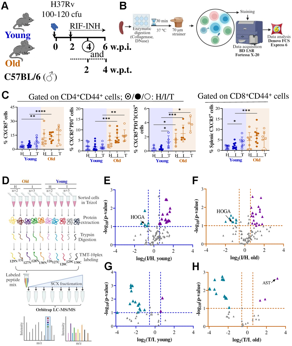
Figure 3.Deregulated splenic CD4+CD44+ T cell proteome in old C57BL/6 mice upon Mtb infection and RIF-INH treatment. (A) Schematic showing Mtb H37Rv aerosol infection in male C57BL/6 mice of two age groups (young: 2-4 and old: 17-19 in months) followed by treatment with RIF-INH starting at 2 w.p.i.. (B) Workflow for immune cell isolation from the spleen, flow cytometry data acquisition and data analysis. (C) Frequencies of CD4+CD44+CXCR5+, CD4+CD44+CXCR5+PD1+ and CD4+CD44+CXCR5+PD1+ICOS+ and CD8+CD44+CXCR5+ cells in the spleen of C57BL/6 mice at 4 w.p.i./2 w.p.t.. Healthy (H; n=3/age group), infected (I; n=6-10/age group) and treated (T; n=3/age group). (D) TMT10plex workflow for CD4+CD44+ T cell proteome analysis (for TMT set-1) by liquid chromatography- mass spectrometry (LC-MS/MS). (E–H) showing volcano plots of splenic CD4+CD44+ T cell proteome showing significantly deregulated proteins (in -log10p-value ≥ 1; log2fold change > |0.58|): (E) Young infected versus healthy, (F) Old infected versus healthy, (G) Young treated versus infected and (H) Old treated versus infected; ▲/▲: up-/down-regulation; HOGA: 4-hydroxy-2-oxoglutarate aldolase; AST: aspartate aminotransferase. Young (2-4 months) mice in blue and old (17-19 months) mice in brown; cfu= colony forming unit; w.p.i.= weeks post infection; p-values: * ≤0.05, ** <0.005, *** <0.0005 and **** <0.0001 at 95% confidence interval by Mann-Whitney test. Data shown as mean ± SD. See also Supplementary Figures 7–10 and Supplementary Tables 1–4.