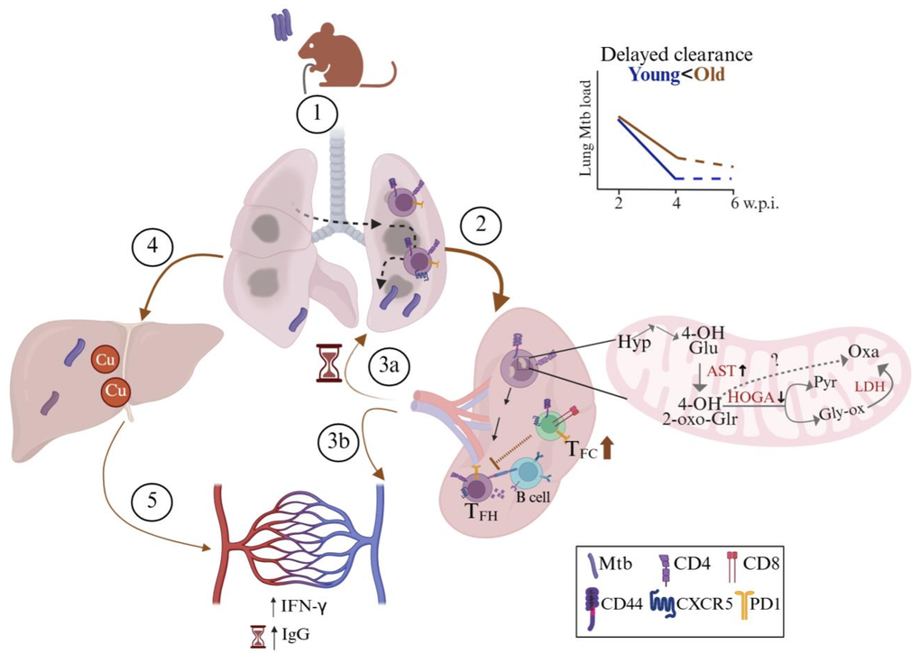
Figure 4.Age-associated mechanisms of delayed Mycobacterium tuberculosis lung clearance in old C57BL/6 mice during early rifampicin-isoniazid treatment. Illustrative summary of the results observed in the study. (1) Following Mtb infection via aerosol challenge, (2) antigen presentation takes place in the spleen. (3a) T cells reach the inflamed lung of old mice. (3b) Increased levels of proinflammatory cytokines: interferon-gamma (IFN-γ) and increased Mtb-specific IgG levels observed at later timepoint in Mtb-infected old mice. (4) Mtb disseminates to the liver via hepatic artery where copper (Cu) was observed to be accumulated (5) alongside increased systemic proinflammatory mediators. Old mice exhibit dysfunctional splenic T follicular helper (TFH : CD4+CD44+CXCR5+PD1+) cells as a virtue of high T follicular cytotoxic cells (TFC : CD8+CD44+CXCR5+PD1+), and leads to delayed lung Mtb clearance at 2 weeks post-treatment. Proteome analysis revealed splenic CD4+CD44+ T cells of old mice showed alterations in mitochondrial proteins (decreased HOGA: 4-hydroxy-2-oxoglutarate aldolase and increased AST), disrupting hydroxyproline (Hyp) degradation and promoting 4-hydroxy-2-oxo-glutarate (4-OH-2-oxo-Glr) accumulation, directing the reaction to oxalate (Oxa) via a pathway independent of lactate dehydrogenase (LDH), pyruvate (Pyr) and glyoxylate (Gly-ox). Created with BioRender.