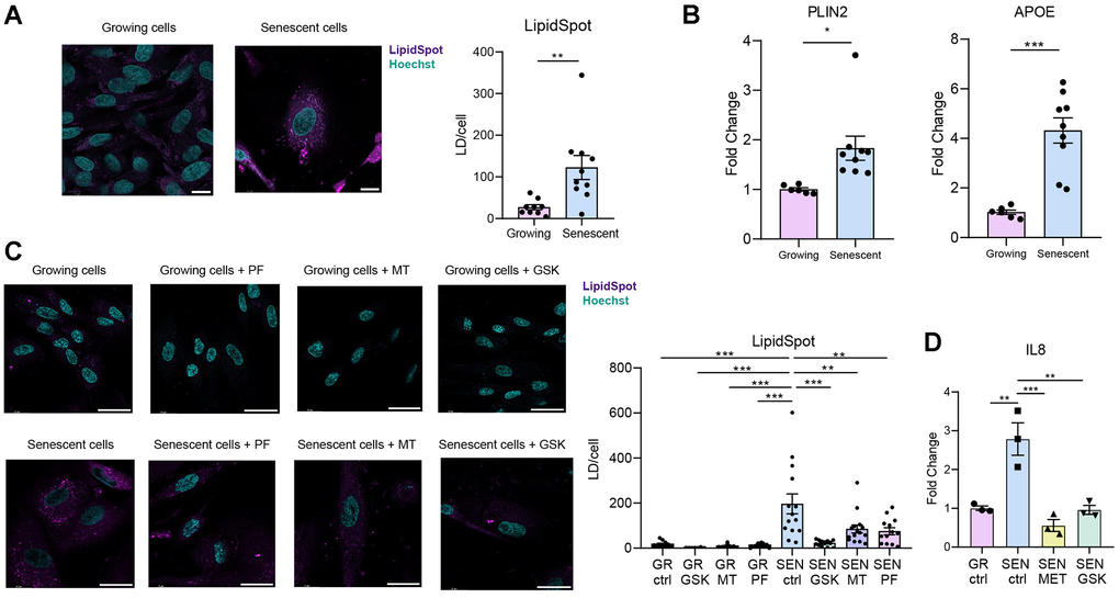
Figure 2.SnCs accumulate lipid droplets (LDs). Senescence was induced in BJ fibroblasts by a 48-hour incubation with 50 μM Etoposide, and cells were analysed one week post treatment [43]. (A) Representative confocal microscopy images and quantification of LDs in growing and senescent BJ fibroblasts, visualized by LipidSpot staining. LDs numbers were counted and normalized to cell count. (B) RT-PCR analysis of the expression of LDs marker genes in senescent BJ fibroblasts compared to growing controls. (C) Representative confocal microscopy images and quantification of LDs in BJ fibroblasts treated with AMPK activators (10 μM for 48 hours) following senescence induction. LDs numbers were counted and normalized to cell count. The significant increase in LDs in senescent BJ cells was reversed by AMPK activators. (D) RT-PCR analysis of the expression of IL8 in senescent BJ fibroblasts non-treated and treated with GSK621 (10 μM) or with Metformin (3mM) compared to growing controls. Statistical significance was assessed using a two-tailed unpaired t-test (*P<0.05, **P<0.005, ***P<0.0005). Data are presented as mean ± SEM. Scale bar = 10 μm.