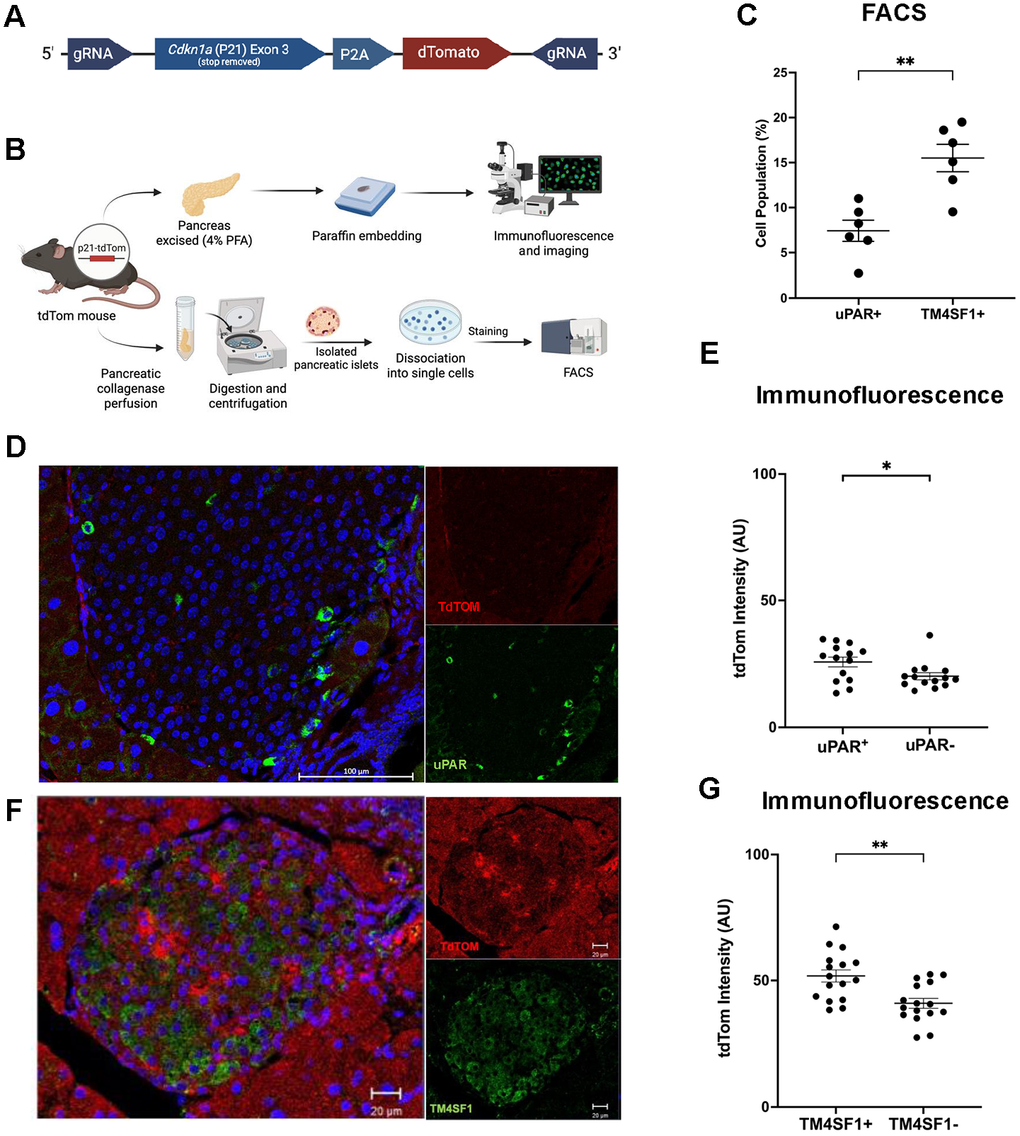
Figure 4.P21-tdTom reporter mouse supports TM4SF1 as a senescent β-cell surface marker. (A) CRISPR/Cas9-mediated knock-in strategy for the p21Cip1-tdTomato reporter mice. (B) Experimental design using P21-tdTom reporter mouse to validate surface markers by flow cytometry (8-18 months of age, n = 6) and immunofluorescence (4-12 months of age, n = 5). (C) Flow cytometry subpopulations of p21-tdTom and uPAR or TM4SF1. Each dot represents islets from a single mouse. (D) Representative confocal image of mouse pancreatic islet from a 53- week-old male uPAR+ mouse stained for tdTom (red) and uPAR (green). Mag bar 100 μm. (E) Immunofluorescence quantification of tdTom expression in uPAR+ vs. uPAR- cells; pancreas from 5 separate mice were evaluated; each dot represents a single cell from the top and bottom 20% of TdTomato intensity. More than 80 cells were measured. (F) Representative confocal image of mouse pancreatic islet from a 23-week-old female tdTom+, stained for tdTom (red) and TM4SF1 (green), indicating senescent β-cell localization. Mag bar 20 μm. (G) Immunofluorescence quantification of tdTom expression in TM4SF1+ vs. TM4SF1- cells pancreas from 5 separate mice were evaluated. Statistically significant higher expression of TdTomato in TM4SF1+ cells; each dot represents a single cell from the top and bottom 20% of TdTom intensity. More than 80 cells were measured. Results are mean ± SEM; *p ≤ 0.05; **p ≤ 0.01.