


Introduction
Lung adenocarcinoma (LUAD) is the most frequent histologic subtype of lung cancer, which is the leading cause of cancer-related mortality worldwide [1]. Early diagnosis of this malignancy is still an optimal opportunity, which is regarded to be a fundamental basis of further therapies, such as targeted and immune-based therapies [2]. Unlike lung squamous cell carcinoma, LUAD mainly occurs and develops within terminal bronchioles and alveoli, which are hardly accessible by bronchoscopy. Therefore, the early-stage of LUAD has been more elusive and difficult to identify [3–5]. A better understanding of the underlying pathogenesis of LUAD would put forward a new insight into discovering the early-stage diagnostic biomarkers to provide an advanced therapeutic strategy for patients with LUAD. Due to the anatomical structure of LUAD, the immune contexture is very complex. Emerging evidence shows that tumor microenvironment impacts LUAD progression and clinical outcome [6]. The tumor microenvironment consists of diverse immune cells, including macrophage and lymphocyte. It has previously been observed that GTPase of immunity-associated proteins (GIMAPs) has been implicated in the regulation of cell survival for lymphomyeloid cells and can be detected highest in lymphoid organs [7]. To date, the GIMAPs cluster are reported on Chromosome 7q36.1, within 300 kb, including eight functional genes and one pseudogene (GIMAP3, also named GIMAP3P). The six genes GIMAP1, GIMAP2, GIMAP4, GIMAP5, GIMAP6, and GIMAP7 encode 33-46 kDa proteins with one GTP-binding domain, whereas GIMAP8 encodes a 75 kDa protein with three GTP-binding domains. GIMAP1-GIMAP5 is a readthrough transcript variant between the neighboring GIMAP1 and GIMAP5, and it encodes a fusion protein that shares sequence identity with each gene product [8]. GIMAPs family members are reported to implicated in the progression of cancer via a regulation of immunologic microenvironment.
Despite growing evidence in understanding the potential importance of GIMAPs in cancer, detailed mechanisms regarding the biological signature of GIMAPs in LUAD await elucidation. Exploring the molecular mechanism of GIMAPs has thus become imperative for advancing diagnosis and therapy of LUAD. In this study, we addressed this problem by a pipeline of systematic analysis (Figure 1), in order to identify the transcriptional and protein expression patterns of GIMAPs family members upon TCGA, Oncomine, and Human Protein Atlas databases. Then we continued to predict biological functions and pathways of GIMAPs as well as their 20 related genes. Subsequently, we analyzed clinical features and prognostic values of GIMAPs family members. Furthermore, we performed an analysis of molecular-disease association between GIMAPs and multiple disorders. The current study links the biological functionality and prognostic value of GIMAPs in LUAD, which will be beneficial to the diagnosis and treatment of lung adenocarcinoma.
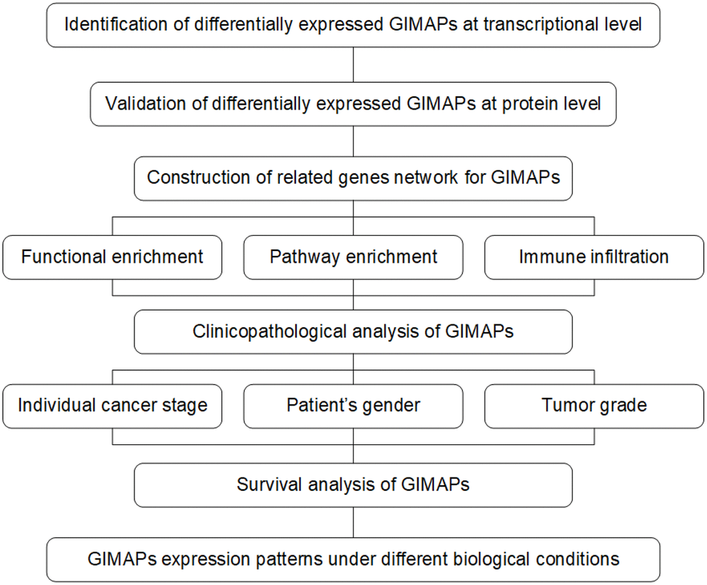
Figure 1. The pipeline of this study based on public databases and platforms.
Results
Absent expression of GIMAPs family members in patients with LUAD
In order to reveal the different potential therapeutic value of GIMAPs in LUAD patients, the mRNA expression of GIMAPs in 20 types of cancers were initially measured and compared to normal tissues by Oncomine database (Figure 2, Table 1). The under- expression of GIMAP1/2/4/5/6/7/8 were found in lung cancer tissues consistently, while neither GIMAP3 nor GIMAP1-GIMAP5 presented available data. In Okayama Lung dataset, GIMAP1 under-expression was found in LUAD tissues compared with normal tissues with a fold change of -3.359 (p=1.37E-13) [9], while Hou observed a 2.848-fold decrease in GIMAP1 mRNA expression in LUAD samples (p=4.73E-18) [10]. Similarly, significant down-regulation of GIMAP4 was also found in Selamat Lung [11], Okayama Lung [9], Hou Lung [10] database. GIMAP5 was also found in Su Lung [12], Selamat Lung [11], Okayama Lung [9], Hou Lung [10] database. GIMAP6 was also found in Landi Lung [13], Su Lung [12], Okayama Lung [9], Hou Lung [10] database. GIMAP7 was also found in Selamat Lung [11], Okayama Lung [9], Hou Lung [10] database. GIMAP8 was also found in Selamat Lung [11] database. However, GIMAP2 was found in the large cell lung carcinoma not in the LUAD with lower mRNA expression. Next, the mRNA expression patterns of GIMAPs were further validated by UALCAN using the resource based on RNA-seq from TCGA database. The mRNA expressions of other GIMAPs members were all found to be significantly down-regulated in LUAD tissues compared to normal samples (Figure 3).
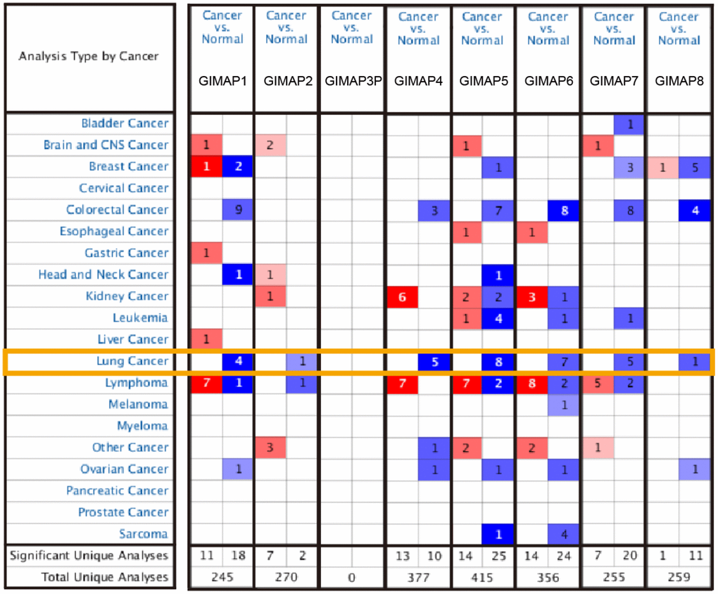
Figure 2. Transcriptional expression of GIMAPs in 20 different types of cancer diseases (Oncomine database). The difference in transcriptional expression was compared by t-test. The threshold statistical parameters were that, p value< 0.0001, fold change = 2, gene rank = 10%, and data type was mRNA.
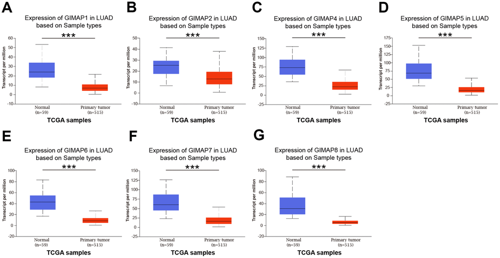
Figure 3. mRNA expression of distinct GIMAPs in LUAD tissues and adjacent normal lung tissues (UALCAN). mRNA expressions of GIMAPs family members were found to be under-expressed in primary LUAD tissues compared to normal samples (A–G). *** p<0.001.
Table 1. Significant changes of GIMAPs expression in transcription level between LUAD and normal lung tissues (ONCOMINE).
| GIMAPs | Types of LUAD VS. Lung | Fold Change | P-value | t-test | Ref | ||||||||||||||||||||||||||||||||||||||||||||||||||||||||||||||||||||||||||||||||||||||||||||||
| GIMAP1 | |||||||||||||||||||||||||||||||||||||||||||||||||||||||||||||||||||||||||||||||||||||||||||||||||||
| Lung Adenocarcinoma | -3.359 | 1.37E-13 | -12.857 | Okayama Lung | |||||||||||||||||||||||||||||||||||||||||||||||||||||||||||||||||||||||||||||||||||||||||||||||
| Lung Adenocarcinoma | -2.848 | 4.73E-18 | -10.665 | Hou Lung | |||||||||||||||||||||||||||||||||||||||||||||||||||||||||||||||||||||||||||||||||||||||||||||||
| GIMAP4 | |||||||||||||||||||||||||||||||||||||||||||||||||||||||||||||||||||||||||||||||||||||||||||||||||||
| Lung Adenocarcinoma | -3.652 | 2.77E-29 | -16.284 | Selamat Lung | |||||||||||||||||||||||||||||||||||||||||||||||||||||||||||||||||||||||||||||||||||||||||||||||
| Lung Adenocarcinoma | -2.921 | 2.13E-08 | -6.564 | Su Lung | |||||||||||||||||||||||||||||||||||||||||||||||||||||||||||||||||||||||||||||||||||||||||||||||
| Lung Adenocarcinoma | -2.02 | 4.1E-09 | -6.72 | Hou Lung | |||||||||||||||||||||||||||||||||||||||||||||||||||||||||||||||||||||||||||||||||||||||||||||||
| GIMAP5 | |||||||||||||||||||||||||||||||||||||||||||||||||||||||||||||||||||||||||||||||||||||||||||||||||||
| Lung Adenocarcinoma | -2.926 | 5.8E-13 | -9.931 | Su Lung | |||||||||||||||||||||||||||||||||||||||||||||||||||||||||||||||||||||||||||||||||||||||||||||||
| Lung Adenocarcinoma | -3.536 | 1.29E-31 | -16.428 | Selamat Lung | |||||||||||||||||||||||||||||||||||||||||||||||||||||||||||||||||||||||||||||||||||||||||||||||
| Lung Adenocarcinoma | -2.41 | 4.38E-12 | -10.856 | Okayama Lung | |||||||||||||||||||||||||||||||||||||||||||||||||||||||||||||||||||||||||||||||||||||||||||||||
| Lung Adenocarcinoma | -2.382 | 4.81E-13 | -8.947 | Hou Lung | |||||||||||||||||||||||||||||||||||||||||||||||||||||||||||||||||||||||||||||||||||||||||||||||
| GIMAP6 | |||||||||||||||||||||||||||||||||||||||||||||||||||||||||||||||||||||||||||||||||||||||||||||||||||
| Lung Adenocarcinoma | -3.755 | 5.99E-25 | -13.546 | Landi Lung | |||||||||||||||||||||||||||||||||||||||||||||||||||||||||||||||||||||||||||||||||||||||||||||||
| Lung Adenocarcinoma | -4.357 | 1.08E-10 | -7.802 | Su Lung | |||||||||||||||||||||||||||||||||||||||||||||||||||||||||||||||||||||||||||||||||||||||||||||||
| Lung Adenocarcinoma | -2.894 | 1.84E-13 | -12.45 | Okayama Lung | |||||||||||||||||||||||||||||||||||||||||||||||||||||||||||||||||||||||||||||||||||||||||||||||
| Lung Adenocarcinoma | -2.906 | 9.91E-14 | -9.448 | Hou Lung | |||||||||||||||||||||||||||||||||||||||||||||||||||||||||||||||||||||||||||||||||||||||||||||||
| GIMAP7 | |||||||||||||||||||||||||||||||||||||||||||||||||||||||||||||||||||||||||||||||||||||||||||||||||||
| Lung Adenocarcinoma | -2.898 | 1.01E-17 | -10.238 | Selamat Lung | |||||||||||||||||||||||||||||||||||||||||||||||||||||||||||||||||||||||||||||||||||||||||||||||
| Lung Adenocarcinoma | -2.495 | 1.21E-11 | -10.53 | Okayama Lung | |||||||||||||||||||||||||||||||||||||||||||||||||||||||||||||||||||||||||||||||||||||||||||||||
| Lung Adenocarcinoma | -2.674 | 4.32E-12 | -8.208 | Hou Lung | |||||||||||||||||||||||||||||||||||||||||||||||||||||||||||||||||||||||||||||||||||||||||||||||
| GIMAP8 | |||||||||||||||||||||||||||||||||||||||||||||||||||||||||||||||||||||||||||||||||||||||||||||||||||
| Lung Adenocarcinoma | -3.113 | 1E-27 | -15.438 | Selamat Lung | |||||||||||||||||||||||||||||||||||||||||||||||||||||||||||||||||||||||||||||||||||||||||||||||
| LUAD: Lung Adenocarcinoma; GIMAP: GTPase of immunity-associated protein. | |||||||||||||||||||||||||||||||||||||||||||||||||||||||||||||||||||||||||||||||||||||||||||||||||||
Furthermore, the protein expression patterns of GIMAPs were examined to describe gene expression from protein level which is closer to the most primitive form of the disease (Figure 4). Firstly, the protein expression patterns of GIMAPs were measured by UALCAN using the resources based on Clinical Proteomic Tumor Analysis Consortium (CPTAC) database. Unfortunately, there was only phosphoprotein not total protein data of GIMAP5, and the protein expression has no significant difference, while other GIMAPs have a high significant difference. Next, the Human Protein Atlas was used to explore the immunohistochemistry images of GIMAPs. GIMAP1/2 proteins were low expressed in normal lung tissues, whereas not expressions of them were observed in LUAD tissue (Figure 5A, 5B). Also, medium protein expressions of GIMAP4/6 were expressed in normal lung tissues, while not or low protein expressions of them was observed in LUAD tissue (Figure 5C, 5D). Moreover, GIMAP7/8 would have high protein expressions in normal lung tissues compared with that in LUAD sample (Figure 5E, 5F).
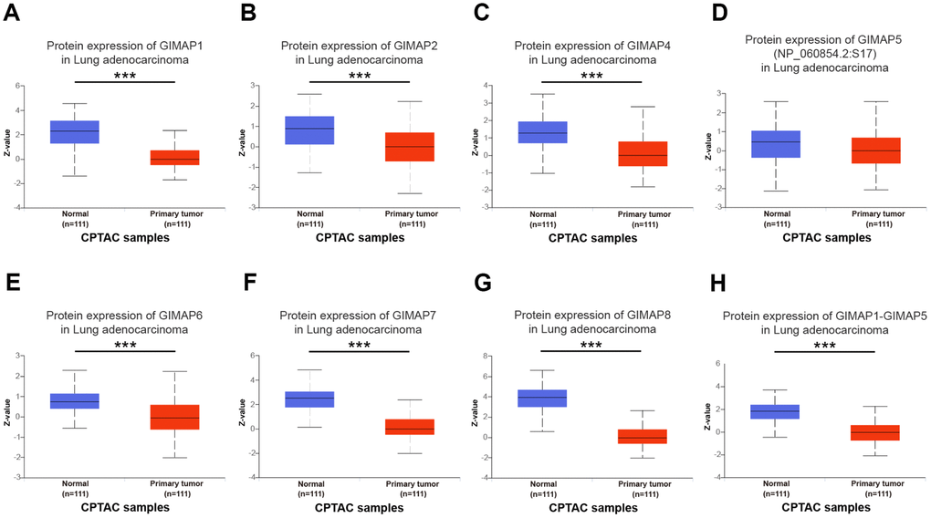
Figure 4. Protein expression of distinct GIMAPs in LUAD tissues and adjacent normal lung tissues (UALCAN). Protein expressions of GIMAPs family members were found to be under-expressed in primary LUAD tissues compared to normal samples (A–H). *** p<0.001.
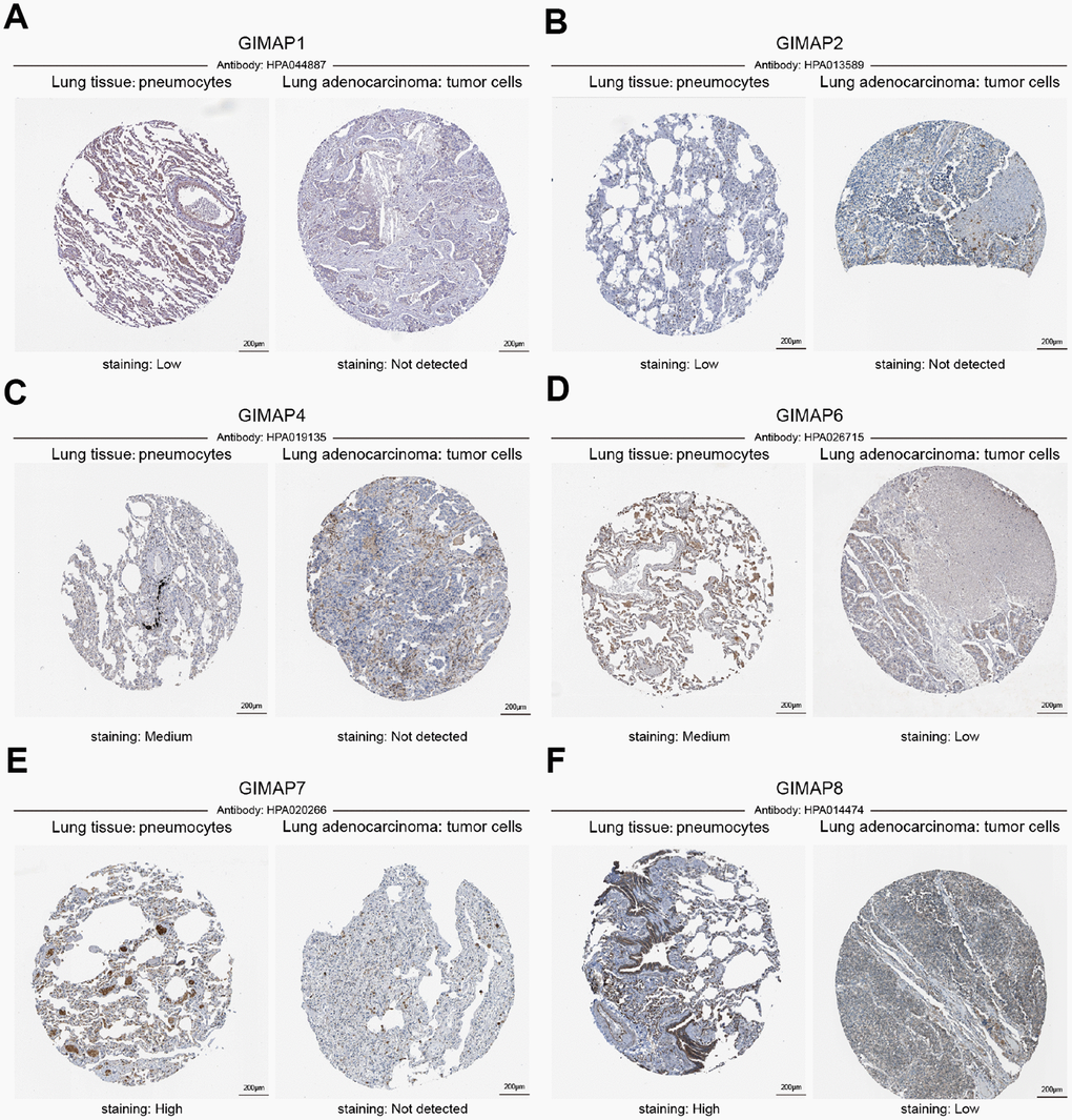
Figure 5. Representative immunohistochemistry images of distinct GIMAPs in LUAD tissues and normal lung tissues (Human Protein Atlas). GIMAP1/2 proteins were low expressed in normal lung tissues, whereas not expressions of them were observed in LUAD tissue (A, B). In addition, medium protein expressions of GIMAP4/6 were expressed in normal lung tissues, while not and low protein expressions of them were observed in LUAD tissue (C, D). Moreover, GIMAP7/8 would have high protein expressions in normal lung tissues compared with the LUAD tissue have not and low protein expressions (E, F).
Generally, all the results above showed that transcriptional and protein expressions of GIMAPs were under-expressed in patients with LUAD.
Predicted functions and pathways of GIMAPs in LUAD patients
To explore the biological function of GIMAPs, we constructed a network of GIMAPs and their 20 related genes by GeneMANIA (Figure 6A). Most of the related genes were in the category named shared protein domains, which means two gene products are linked if they have the same protein domain.
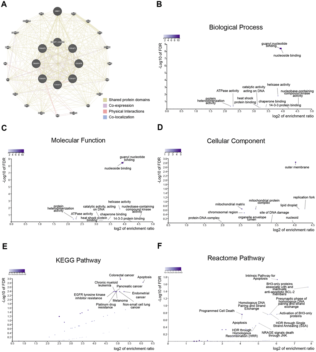
Figure 6. Predicted functions and pathways of GIMAPs and their 20 related genes in LUAD patients (GeneMANIA and WebGestalt). Network of GIMAPs and their 20 related genes was constructed (A). GO functional enrichment analysis predicted three main functions of GIMAPs and their 20 related genes, including biological process, cellular components and molecular functions (B–D). KEGG and Reactome pathway analysis on GIMAPs and their 20 related genes was shown at figure (E, F).
Moreover, GO functions and pathways of GIMAPs and their 20 related genes were analyzed by WEB-based Gene SeT AnaLysis Toolkit (WebGestalt). The biological processes such as GO:0051052 (regulation of DNA metabolic process), GO:0038034 (signal transduction in absence of ligand), GO:0090559 (regulation of membrane permeability), and GO:0006260 (DNA replication) were remarkably regulated by the GIMAPs in LUAD (Figure 6B and Table 2). Cellular components, including GO:0019867 (outer membrane), GO:0005759 (mitochondrial matrix), GO:0005657 (replication fork), GO:0005811 (lipid droplet) and GO:0098798 (mitochondrial protein complex) were significantly associated with the GIMAPs alterations (Figure 6C). Besides, GIMAPs also prominently involved in the molecular functions, such as GO:0001882 (nucleoside binding), GO:0019001 (guanyl nucleotide binding), GO:0004386 (helicase activity), GO:0019205 (nucleobase-containing compound kinase activity) and GO:0140097 (catalytic activity, acting on DNA) (Figure 6D).
Table 2. The top 5 enriched GO terms of the GIMAPs and their 20 related genes in LUAD patients (WebGestalt).
| Category | GO ID | GO term | Count | FDR | Log P | Ratio |
| Biological Process | GO:0051052 | regulation of DNA metabolic process | 405 | 0.0048132 | -5.25 | 12.188 |
| Biological Process | GO:0038034 | signal transduction in absence of ligand | 69 | 0.031887 | -4.12 | 35.771 |
| Biological Process | GO:0090559 | regulation of membrane permeability | 79 | 0.031887 | -3.95 | 31.243 |
| Biological Process | GO:0006260 | DNA replication | 268 | 0.04701 | -3.58 | 12.279 |
| Biological Process | GO:0071887 | leukocyte apoptotic process | 107 | 0.04701 | -3.56 | 23.067 |
| Cellular Component | GO:0019867 | outer membrane | 206 | 0.024847 | -3.84 | 13.979 |
| Cellular Component | GO:0005759 | mitochondrial matrix | 462 | 0.18341 | -2.52 | 6.2331 |
| Cellular Component | GO:0005657 | replication fork | 69 | 0.18341 | -2.4 | 20.867 |
| Cellular Component | GO:0005811 | lipid droplet | 77 | 0.18341 | -2.31 | 18.699 |
| Cellular Component | GO:0098798 | mitochondrial protein complex | 266 | 0.18341 | -2.27 | 8.1195 |
| Molecular Function | GO:0001882 | nucleoside binding | 384 | 0.914081 | -12.15 | 16.745 |
| Molecular Function | GO:0019001 | guanyl nucleotide binding | 390 | 1.19624 | -12.07 | 16.487 |
| Molecular Function | GO:0004386 | helicase activity | 150 | 3.348849 | -3.81 | 14.289 |
| Molecular Function | GO:0019205 | nucleobase-containing compound kinase activity | 46 | 4.30035 | -2.48 | 23.297 |
| Molecular Function | GO:0140097 | catalytic activity, acting on DNA | 184 | 11.86587 | -2.33 | 8.7363 |
In KEGG analysis, these pathways including hsa01521 (EGFR tyrosine kinase inhibitor resistance), hsa05210 (colorectal cancer), hsa04210 (apoptosis), hsa05014 (amyotrophic lateral sclerosis) and hsa05213 (endometrial cancer) were associated with the functions of GIMAPs in LUAD (Figure 6E and Table 3). While in the Reactome analysis, the most significantly enriched terms were involved in R-HSA-109606 (intrinsic pathway for apoptosis), R-HSA-111453 (BH3-only proteins associate with and inactivate anti-apoptotic BCL-2 members), R-HSA-114452 (activation of BH3-only proteins), R-HSA-109581 (apoptosis), and R-HSA-5357801 (programmed cell death) (Figure 6F and Table 4).
Table 3. The top 5 enriched KEGG pathway terms of the GIMAPs and their 20 related genes in LUAD patients (WebGestalt).
| Pathway ID | Pathway name | Count | FDR | Log P | Ratio |
| hsa01521 | EGFR tyrosine kinase inhibitor resistance | 79 | 0.019198 | -4.04 | 31.515 |
| hsa05210 | Colorectal cancer | 86 | 0.019198 | -3.93 | 28.95 |
| hsa04210 | Apoptosis | 136 | 0.049746 | -3.34 | 18.306 |
| hsa04215 | Apoptosis | 33 | 0.05448 | -3.17 | 50.296 |
| hsa05014 | Amyotrophic lateral sclerosis | 51 | 0.10407 | -2.8 | 32.545 |
Table 4. The top 5 Reactome terms of the GIMAPs and their 20 related genes in LUAD patients (WebGestalt).
| Pathway ID | Pathway name | Count | FDR | Log P | Ratio |
| R-HSA-109606 | Intrinsic Pathway for Apoptosis | 44 | 0.018834 | -4.96 | 65.417 |
| R-HSA-111453 | BH3-only proteins associate with and inactivate anti-apoptotic BCL-2 members | 9 | 0.030598 | -4.45 | 213.21 |
| R-HSA-114452 | Activation of BH3-only proteins | 30 | 0.1769 | -3.37 | 63.964 |
| R-HSA-109581 | Apoptosis | 169 | 0.1769 | -3.22 | 17.032 |
| R-HSA-5357801 | Programmed Cell Death | 172 | 0.1769 | -3.2 | 16.735 |
After GO and pathway analyzing, immune infiltration of GIMAPs was estimated by Tumor IMmune Estimation Resource (TIMER). The scatterplots of GIMAPs showed a purity-corrected value and statistical significance. All GIMAPs had negative associations with tumor purity, which means these genes were highly involved in the progression of immune infiltration in LUAD (Figure 7 and Table 5).
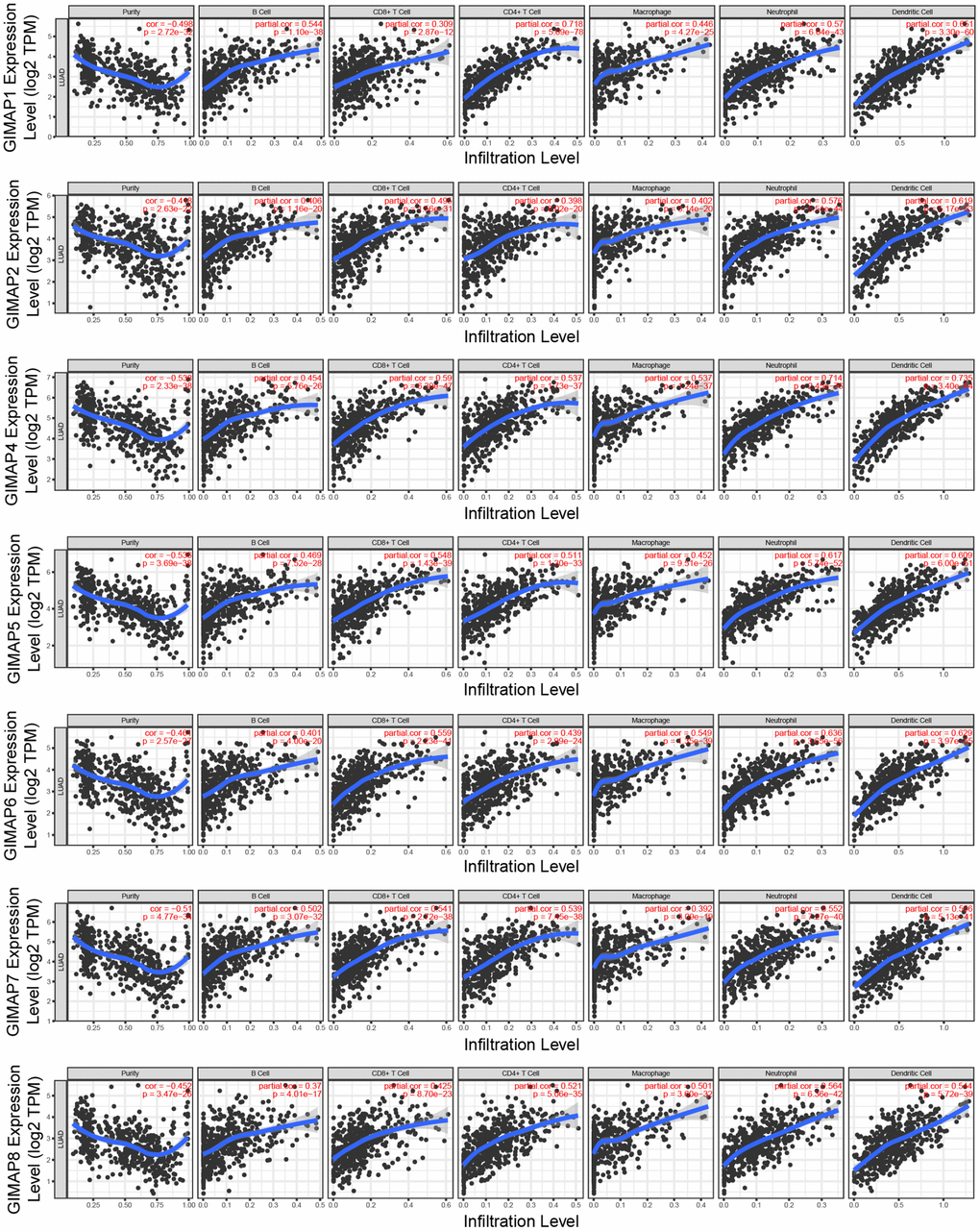
Figure 7. Immune infiltration analysis of GIMAPs family members (TIMER). The purity-corrected value of all GIMAPs were negative, that means these genes were highly expressed in the microenvironment.
Table 5. Correlation coefficient of immune infiltration level with GIMAPs expression level (TIMER).
| Purity | B cell | CD8+ T cell | CD4+ T cell | Macrophage | Neutrophil | Dendritic cell | |
| GIMAP1 | -0.4989 (p = 2.72e-32) | 0.544 (p = 1.10e-38) | 0.309 (p = 2.87e-12) | 0.718 (p = 5.69e-78) | 0.446 (p = 4.27e-25) | 0.57 (p = 6.04e-43) | 0.651 (p = 3.30e-60) |
| GIMAP2 | -0.418 (p = 2.63e-22) | 0.406 (p = 1.16e-20) | 0.495 (p = 1.56e-31) | 0.398 (p = 8.02e-20) | 0.402 (p = 3.14e-20) | 0.576 (p = 5.51e-44) | 0.619 (p = 5.17e-53) |
| GIMAP4 | -0.538 (p = 2.33e-38) | 0.454 (p = 5.76e-26) | 0.59 (p = 6.29e-47) | 0.537 (p = 1.73e-37) | 0.537 (p = 1.24e-37) | 0.714 (p = 2.45e-76) | 0.735 (p = 3.40e-84) |
| GIMAP5 | -0.538 (p = 3.69e-38) | 0.469 (p = 7.52e-28) | 0.548 (p = 1.43e-39) | 0.511 (p = 1.30e-33) | 0.452 (p = 9.51e-26) | 0.617 (p = 5.74e-52) | 0.609 (p = 6.00e-51) |
| GIMAP6 | -0.461 (p = 2.57e-27) | 0.401 (p = 4.00e-20) | 0.559 (p = 2.23e-41) | 0.439 (p = 2.99e-24) | 0.549 (p = 1.35e-39) | 0.636 (p = 3.65e-56) | 0.629 (p = 3.97e-55) |
| GIMAP7 | -0.51 (p = 4.77e-34) | 0.502 (p = 3.07e-32) | 0.541 (p = 2.72e-38) | 0.539 (p = 7.45e-38) | 0.392 (p = 3.09e-19) | 0.552 (p = 7.27e-40) | 0.566 (p = 5.13e-14) |
| GIMAP8 | -0.452 (p = 3.47e-26) | 0.37 (p = 4.01e-17) | 0.425 (p = 8.70e-23) | 0.521 (p = 5.06e-35) | 0.501 (p = 3.60e-32) | 0.564 (p = 6.36e-42) | 0.544 (p = 5.72e-39) |
Taken together, the results showed that GIMAPs played a potential promotive role in the regulation of the DNA metabolic process and involved in apoptosis, as well as immune infiltration.
Association of transcriptional and protein expression of GIMAPs family members with clinicopathological features of LUAD patients
Further, we analyzed a relationship between GIMAPs’ expression with the clinicopathological characteristics of LUAD patients by UALCAN, including individual cancer stage, patient’s gender, and tumor grade. The mRNA expression of GIMAPs based on RNA-seq was from TCGA database and protein expression from CPTAC. The mRNA expressions of GIMAPs were remarkably correlated with patients’ individual cancer stages, and patients who were in more advanced cancer stages tended to express lower mRNA expression of GIMAPs (Figure 8). Similarly, mRNA expressions of GIMAPs were significantly related to patient’s gender, and male patients tended to express lower mRNA expression (Figure 9). Besides that, protein expression of GIMAPs were significantly related to tumor grade, as tumor grade increased, the protein expression of GIMAPs tended to be higher (Figure 10). In short, these results suggested that 8 GIMAPs family members were significantly associated with clinicopathological features in LUAD patients.
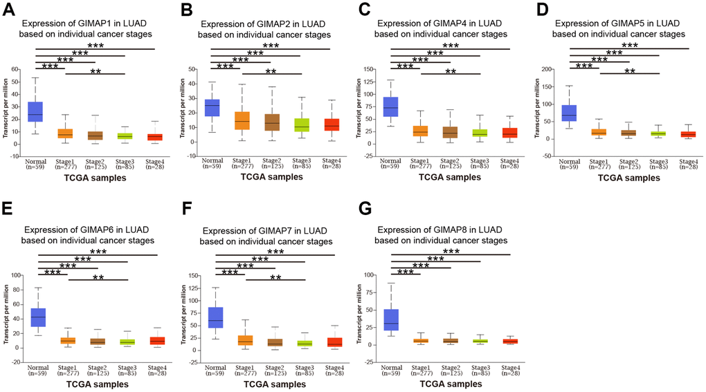
Figure 8. Relationship between mRNA expression of distinct GIMAPs and individual cancer stages of LUAD patients (UALCAN). mRNA expressions of GIMAPs family members were remarkably correlated with patients’ individual cancer stages, patients who were in more advanced cancer stages tended to express lower mRNA expression of GIMAPs (A–G). **p<0.01, ***p<0.001.
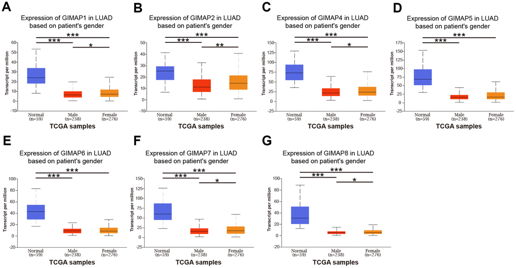
Figure 9. Relationship between mRNA expression of distinct GIMAPs and patient’s gender of LUAD (UALCAN). mRNA expressions of GIMAPs family members were remarkably correlated with patient’s gender, male patients tended to express lower mRNA expression (A–G). *p<0.05, **p<0.01, ***p<0.001.
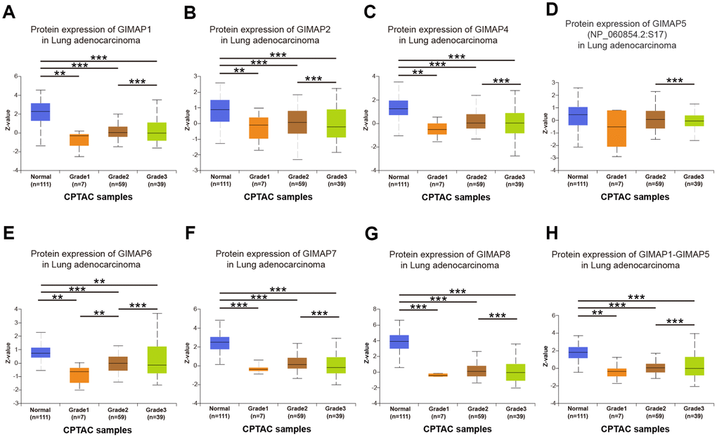
Figure 10. Association of protein expression of distinct GIMAPs with tumor grades of LUAD patients (UALCAN). protein expression of GIMAPs were significantly related to tumor grade, as tumor grade increased, the protein expression of GIMAPs tended to be higher (A–H). **p<0.01, ***p<0.001.
Prognostic value of mRNA expression of GIMAPs in LUAD patients
To analyze the prognostic value of GIMAPs in LUAD patients, we carried out a prognostic evaluation for the mRNA expression of GIMAPs in LUAD patients by GEPIA. As the results showed, mRNA expressions of all GIMAPs family members were significantly associated with LUAD patients’ prognosis, higher mRNA expression of GIMAP1 (HR=0.68, and p=0.0098), GIMAP2 (HR=0.69, and p=0.016), GIMAP4 (HR=0.7, and p=0.017), GIMAP5 (HR=0.63, and p=0.0026), GIMAP6 (HR=0.58, and p=0.00035), GIMAP7 (HR=0.57, and p=0.00018), GIMAP8 (HR=0.57, and p=0.00023), GIMAP1-GIMAP5 (HR=0.63, and p=0.0029) were significantly associated with longer overall survival of LUAD patients (Figure 11). These results indicated that higher mRNA expressions of GIMAPs could increase the survival time of LUAD patients and they may be exploited as potentially useful biomarkers to predict the LUAD patients’ survival.
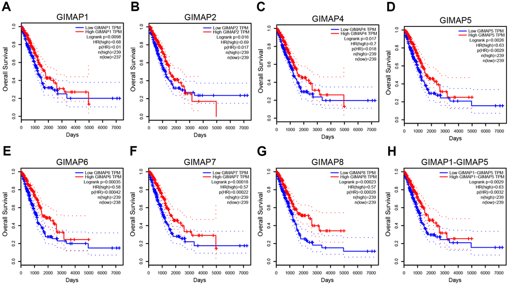
Figure 11. Prognostic value of mRNA expression of distinct GIMAPs in LUAD patients (GEPIA). mRNA expressions of all the GIMAPs family members were significantly associated with LUAD patients’ prognosis, higher mRNA expression of GIMAPs were significantly associated with longer overall survival of LUAD patients (A–H).
The molecular-disease association of GIMAPs with multiple diseases
After analyzing the prognostic value of GIMAPs in LUAD patients, we further explore the relationship of GIMAPs with various diseases by the Open Targets Platform. GIMAP1/4/6/7/8 were predicted to have a higher association with LUAD filtered by RNA expression in this study, while GIMAP2 was predicted to have an association with lung carcinoma and GIMAP2 was predicted to have an association with sepsis (Figure 12). All in a word, GIMAPs had a close relationship with LUAD and they may be predicted as potential therapeutic targets for LUAD patients.
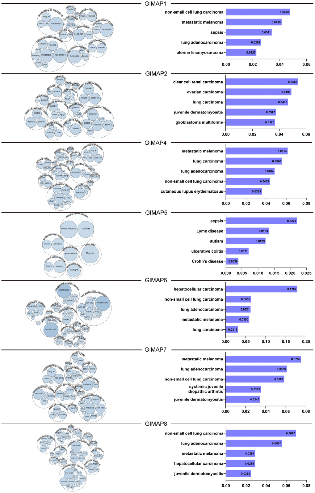
Figure 12. Relationship between GIMAPs with LUAD among the different disease (the Open Targets Platform). GIMAPs had a close relationship with LUAD and could be determined and visualized. The bubbles color and bubbles size represent the association score, and the top 5 diseases with a high score value are listed at right.
Discussion
In the tumor microenvironment, aberrant lymphocyte immune infiltration has been found to take part in the development and progression of LUAD [7]. Being important factors of regulation complexes for immune infiltration, dysregulation expression of GIMAPs family are implicated in the development of LUAD [14]. However, distinct roles of GIMAPs family members in LUAD remained to be elucidated. In this study, the transcriptional and protein expression of GIMAPs family members in LUAD were identified based on distinct public databases. Furthermore, the mRNA expression of GIMAPs was significantly associated with patients’ individual cancer stages and patient’s gender in LUAD patients, while the protein expression of GIMAPs was significantly associated with tumor grades in LUAD patients. Moreover, higher mRNA expressions of GIMAPs significantly associated with longer OS in LUAD patients. Finally, as in the target-disease relationship, GIMAPs had a close relationship with cell proliferation disorders, especially LUAD.
The functions enrichment of GIMAPs and their 20 related genes in LUAD patients were analyzed and our results showed that biological processes such as GO:0051052 (regulation of DNA metabolic process), cellular components such as GO:005657 (replication fork), and molecular functions such as GO:0001882 (nucleoside binding) were remarkably regulated by the GIMAPs and their 20 related genes in LUAD. Mechanistic studies in vitro reveal that impairment of GIMAP5-deficient TH cell differentiation is associated with increased DNA damage, which could be controlled by TGF-βin GIMAP5-deficient CD4+ T cells to induce TH17 polarization [15]. Previous in vivo study showed that GIMAP3/5 modified mitochondrial DNA in a heteroplasmic mouse model [16], which was consistent with our findings that GIMAPs family members influenced DNA metabolism in either mitochondria or nucleus.
In this study, KEGG pathways enrichment such as hsa05210 (colorectal cancer) and hsa05213 (endometrial cancer) indicated that GIMAPs mediated progression of various malignancy not only lung cancer. It was reported that the downregulation of the mRNA and protein expression levels of GIMAP5 and GIMAP6 in the tumor tissues and blood of patients with hepatocellular carcinoma. That suggested GIMAP5 and GIMAP6 could involve in the pathogenesis of hepatocellular carcinoma [17]. GIMAP6 had been reported as a one of the hub genes in associated with the pathogenesis of LUAD using the data from the Gene Expression Omnibus (GEO) database. GIMAP6 was expressed at lower levels in the tumor samples with a better prognosis [18], which is the same with our study. GIMAP7 was associated with the infiltration levels of immunocytes, and the inhibition of GIMAP7 would down-regulated the level of FOXO1 expression in pancreatic adenocarcinoma [19]. Mechanistically, GIMAP6 and GIMAP8 showed a reduction of gene expression in the tumors and GIMAP8 mRNA level was abnormally elevated in the adjacent nontumor tissues as compared, which was confirmed by quantitative PCR assays [14]. These studies accord with our implication that GIMAPs family members may play potent roles on tumorigenesis and progression of cancer.
According to Reactome analysis of R-HSA-109606/10958/5357801(apoptosis), our findings exhibited that GIMAPs were involved in regulation of apoptosis. Previous evidence has established that GIMAPs family members are important in regulating apoptosis in cancer cells [17]. GIMAP3 and GIMAP5 proteins seem to play essential roles in T cell homeostasis, via notably ability to improve the activity of Bcl-2 family members [20, 21]. Studies revealed GIMAP4 regulated apoptosis of T-cell by intrinsic stimuli downstream of caspase-3 activation and phosphatidylserine exposure. And the execution of programmed cell death directly correlates with the phosphorylation status of GIMAP4 [22]. Datta P et al. also reported that GIMAP1 ablation is accompanied by activation of the extrinsic apoptotic pathway [23]. Given that pathway and Reactome analyses of GIMAPs, it is likely that GIMAPs are involved in molecular pathogenesis of cancer via regulation of apoptosis. However, this hypothesis needs to be tested.
Among GIMAPs family members, GIMAP1/2/4/7/8 provided a significant difference between genders, which suggested a potential role of GIMAPs with sex hormone. Prior research based on RNA-seq noted a clue between GIMAPs and hormone [24]. But little is known regarding the depth molecular mechanism underlying a relationship between GIMAPs and hormone. In addition, clinical traits of LUAD patients were used to assess the clinical features of GIMAPs. We found that GIMAPs expression laid out a significance between groups with different stages as well as grades. From our findings of these clinical characteristics, GIMAPs family members were significantly associated with stage and grade of LUAD. The independent prognostic value of GIMAPs in LUAD pleas for a further understanding of mechanism by which GIMAPs family members drive tumorigenesis and development of lung adenocarcinoma.
Results from our study showed that lack of mRNA and protein expression was found in all the GIMAPs family members in LUAD, which suggested that lack of GIMAPs expressions would be one of predictors in LUAD. However, there is an academic report presented that aberrant activation of the GIMAP enhancer contributed to regulate the transcription factor TAL1/SCL, which was one of the most prevalent oncogenes in T-cell acute lymphoblastic leukemia [25]. Hence a further investigation would be conducted for exploring the molecular mechanism underlying a biological function of GIMAPs family members.
Obviously, there were some limitations in this study. First, all the data analyzed was based on bioinformatics from the online databases, further in vivo and in vitro studies are required to verify the biological functions of these findings. Second, we did not explore the underlying mechanisms of distinct GIMAPs in LUAD. Further fully designed experiments and analyses are worth to reveal the detailed mechanism between GIMAPs and LUAD. Finally, this study was only a retrospective study, and further prospective results are needed to support each other.
In conclusion, our results showed that under expression of GIMAPs members was found in the LUAD patients based on distinct public databases. GIMAPs were significantly associated with individual cancer stage, patient’s gender, and tumor grade. Besides, GIMAPs family members accompanied with immune infiltration of LUAD. Moreover, high mRNA expressions of GIMAPs were significantly related with longer OS in LUAD patients. All in a word, GIMAPs may become the potential diagnostic biomarkers and predicted as useful survival biomarkers for the LUAD patients.
Materials and Methods
Identification of differentially expressed GIMAPs at transcriptional level
Oncomine (https://www.oncomine.org/resource/login.html) provides robust, peer-reviewed analysis methods and a powerful set of analysis functions to compute gene expression signatures [26]. In this study, Oncomine database is used to identify the transcriptional expressions of GIMAPs family members in LUAD tissues with their corresponding adjacent normal control samples. The data in our study was compared by t-test and the threshold statistical parameters were that, p value < 0.0001, fold change = 2, gene rank = 10%, and data type was mRNA.
UALCAN (http://ualcan.path.uab.edu) is a comprehensive and interactive web resource based on RNA-seq of 26 cancer types from TCGA database [27]. To determine the reliability of the differential expressed data, UALCAN database was chosen to further verify. In this study, the mRNA expressions of different GIMAPs between LUAD tissues with normal tissues were performed in the TCGA-LUAD dataset with a statically significant p < 0.001.
Identification of differentially expressed GIMAPs at protein level
Besides the transcriptional analysis from TCGA, UALCAN database could provide protein expression analysis using data from CPTAC Confirmatory/Discovery dataset for LUAD [28]. The proteome-based subtypes could be observed in external cancer proteomic datasets. The protein expressions of GIMAPs family members between LUAD tissues with normal tissues were performed with a statically significant p < 0.001.
The Human Protein Atlas (https://www.proteinatlas.org) is a website that contains protein expression data for near 20 highly common kinds of cancers based on representative immunohistochemistry data [29]. In order to avoid the result bias caused by data from a single database source, the Human Protein Atlas was applied to identify the tumor-type specific proteins expression patterns of GIMAPs that are differentially expressed in LUAD patients. The immunohistochemistry images of GIMAPs expressed in LUAD and normal tissues could be directly visualized.
Clinicopathological analysis of GIMAPs in LUAD
Furthermore, UALCAN was used to analyze the association between the mRNA or protein expressions of GIMAPs in LUAD tissues with their clinicopathologic parameters such as individual cancer stage, patient’s gender, and tumor grade. The results could be got directly by selecting the clinicopathological grouping options integrated into the UALCAN database. In particular, only the tumor group could be divided into different clinicopathological groups. The statically significant p is less than 0.001.
Survival analysis of GIMAPs
The prognostic value of mRNA expression of distinct GIMAPs in LUAD was analyzed by using GEPIA (http://gepia.cancer-pku.cn/index.html) [33]. which could analyze the RNA sequencing expression data of 9,736 tumors and 8,587 normal samples from the TCGA and the GTEx projects. In this study, patients with LUAD were divided into high and low expression groups based on median values of mRNA expression. The statically significant difference was considered when a p-value < 0.05.
GIMAPs expression patterns under different biological conditions
The Open Targets Platform (https://www.targetvalidation.org) is a comprehensive and powerful data integration for accessing and visualizing potential drug targets related to diseases [34]. To further explore the relationship between GIMAPs with LUAD among multiple diseases, the Open Targets Platform was used to show the available evidence for GIMAPs-disease associations. Data type was filtered by mRNA expression in this study. All methods and Strategies above could be seen in the flow chart below (Figure 1).
Author Contributions
SD wrote this manuscript. ZZ and XL contributed to organizing data from various databases and completing analyses. QZ designed and edited the final version of figures and tables. SX and ML designed the study and corrected the manuscript. All authors read and approved the final manuscript.
Acknowledgments
Authors acknowledge the Helixlife for the introduction of bioinformatics methods.
Conflicts of Interest
The authors declare that they have no conflicts of interests.
References
- 1. Lee JJ, Park S, Park H, Kim S, Lee J, Lee J, Youk J, Yi K, An Y, Park IK, Kang CH, Chung DH, Kim TM, et al. Tracing oncogene rearrangements in the mutational history of lung adenocarcinoma. Cell. 2019; 177:1842–57.e21. https://doi.org/10.1016/j.cell.2019.05.013 [PubMed]
- 2. Kadara H, Kabbout M, Wistuba II. Pulmonary adenocarcinoma: a renewed entity in 2011. Respirology. 2012; 17:50–65. https://doi.org/10.1111/j.1440-1843.2011.02095.x [PubMed]
- 3. Lee HY, Choi YL, Lee KS, Han J, Zo JI, Shim YM, Moon JW. Pure ground-glass opacity neoplastic lung nodules: histopathology, imaging, and management. AJR Am J Roentgenol. 2014; 202:W224–33. https://doi.org/10.2214/AJR.13.11819 [PubMed]
- 4. Travis WD, Brambilla E, Nicholson AG, Yatabe Y, Austin JH, Beasley MB, Chirieac LR, Dacic S, Duhig E, Flieder DB, Geisinger K, Hirsch FR, Ishikawa Y, et al, and WHO Panel. The 2015 world health organization classification of lung tumors: impact of genetic, clinical and radiologic advances since the 2004 classification. J Thorac Oncol. 2015; 10:1243–60. https://doi.org/10.1097/JTO.0000000000000630 [PubMed]
- 5. Yatabe Y, Borczuk AC, Powell CA. Do all lung adenocarcinomas follow a stepwise progression? Lung Cancer. 2011; 74:7–11. https://doi.org/10.1016/j.lungcan.2011.05.021 [PubMed]
- 6. Remark R, Becker C, Gomez JE, Damotte D, Dieu-Nosjean MC, Sautès-Fridman C, Fridman WH, Powell CA, Altorki NK, Merad M, Gnjatic S. The non-small cell lung cancer immune contexture. A major determinant of tumor characteristics and patient outcome. Am J Respir Crit Care Med. 2015; 191:377–90. https://doi.org/10.1164/rccm.201409-1671PP [PubMed]
- 7. Nitta T, Takahama Y. The lymphocyte guard-IANs: regulation of lymphocyte survival by IAN/GIMAP family proteins. Trends Immunol. 2007; 28:58–65. https://doi.org/10.1016/j.it.2006.12.002 [PubMed]
- 8. Krücken J, Schroetel RM, Müller IU, Saïdani N, Marinovski P, Benten WP, Stamm O, Wunderlich F. Comparative analysis of the human gimap gene cluster encoding a novel GTPase family. Gene. 2004; 341:291–304. https://doi.org/10.1016/j.gene.2004.07.005 [PubMed]
- 9. Okayama H, Kohno T, Ishii Y, Shimada Y, Shiraishi K, Iwakawa R, Furuta K, Tsuta K, Shibata T, Yamamoto S, Watanabe S, Sakamoto H, Kumamoto K, et al. Identification of genes upregulated in ALK-positive and EGFR/KRAS/ALK-negative lung adenocarcinomas. Cancer Res. 2012; 72:100–11. https://doi.org/10.1158/0008-5472.CAN-11-1403 [PubMed]
- 10. Hou J, Aerts J, den Hamer B, van Ijcken W, den Bakker M, Riegman P, van der Leest C, van der Spek P, Foekens JA, Hoogsteden HC, Grosveld F, Philipsen S. Gene expression-based classification of non-small cell lung carcinomas and survival prediction. PLoS One. 2010; 5:e10312. https://doi.org/10.1371/journal.pone.0010312 [PubMed]
- 11. Selamat SA, Chung BS, Girard L, Zhang W, Zhang Y, Campan M, Siegmund KD, Koss MN, Hagen JA, Lam WL, Lam S, Gazdar AF, Laird-Offringa IA. Genome-scale analysis of DNA methylation in lung adenocarcinoma and integration with mRNA expression. Genome Res. 2012; 22:1197–211. https://doi.org/10.1101/gr.132662.111 [PubMed]
- 12. Su LJ, Chang CW, Wu YC, Chen KC, Lin CJ, Liang SC, Lin CH, Whang-Peng J, Hsu SL, Chen CH, Huang CY. Selection of DDX5 as a novel internal control for Q-RT-PCR from microarray data using a block bootstrap re-sampling scheme. BMC Genomics. 2007; 8:140. https://doi.org/10.1186/1471-2164-8-140 [PubMed]
- 13. Landi MT, Dracheva T, Rotunno M, Figueroa JD, Liu H, Dasgupta A, Mann FE, Fukuoka J, Hames M, Bergen AW, Murphy SE, Yang P, Pesatori AC, et al. Gene expression signature of cigarette smoking and its role in lung adenocarcinoma development and survival. PLoS One. 2008; 3:e1651. https://doi.org/10.1371/journal.pone.0001651 [PubMed]
- 14. Shiao YM, Chang YH, Liu YM, Li JC, Su JS, Liu KJ, Liu YF, Lin MW, Tsai SF. Dysregulation of GIMAP genes in non-small cell lung cancer. Lung Cancer. 2008; 62:287–94. https://doi.org/10.1016/j.lungcan.2008.03.021 [PubMed]
- 15. Patterson AR, Bolcas P, Lampe K, Cantrell R, Ruff B, Lewkowich I, Hogan SP, Janssen EM, Bleesing J, Khurana Hershey GK, Hoebe K. Loss of GTPase of immunity-associated protein 5 (Gimap5) promotes pathogenic CD4+ T-cell development and allergic airway disease. J Allergy Clin Immunol. 2019; 143:245–57.e6. https://doi.org/10.1016/j.jaci.2018.10.018 [PubMed]
- 16. Jokinen R, Lahtinen T, Marttinen P, Myöhänen M, Ruotsalainen P, Yeung N, Shvetsova A, Kastaniotis AJ, Hiltunen JK, Öhman T, Nyman TA, Weiler H, Battersby BJ. Quantitative changes in Gimap3 and Gimap5 expression modify mitochondrial DNA segregation in mice. Genetics. 2015; 200:221–35. https://doi.org/10.1534/genetics.115.175596 [PubMed]
- 17. Huang Z, Zhang W, Gao C, Ji B, Chi X, Zheng W, Wang HL. Dysregulation of GTPase IMAP family members in hepatocellular cancer. Mol Med Rep. 2016; 14:4119–23. https://doi.org/10.3892/mmr.2016.5764 [PubMed]
- 18. Yu DH, Huang JY, Liu XP, Ruan XL, Chen C, Hu WD, Li S. Effects of hub genes on the clinicopathological and prognostic features of lung adenocarcinoma. Oncol Lett. 2020; 19:1203–14. https://doi.org/10.3892/ol.2019.11193 [PubMed]
- 19. Meng Z, Ren D, Zhang K, Zhao J, Jin X, Wu H. Using ESTIMATE algorithm to establish an 8-mRNA signature prognosis prediction system and identify immunocyte infiltration-related genes in pancreatic adenocarcinoma. Aging (Albany NY). 2020; 12:5048–70. https://doi.org/10.18632/aging.102931 [PubMed]
- 20. Ciucci T, Bosselut R. Gimap and T cells: a matter of life or death. Eur J Immunol. 2014; 44:348–51. https://doi.org/10.1002/eji.201344375 [PubMed]
- 21. Yano K, Carter C, Yoshida N, Abe T, Yamada A, Nitta T, Ishimaru N, Takada K, Butcher GW, Takahama Y. Gimap3 and Gimap5 cooperate to maintain t-cell numbers in the mouse. Eur J Immunol. 2014; 44:561–72. https://doi.org/10.1002/eji.201343750 [PubMed]
- 22. Schnell S, Démollière C, van den Berk P, Jacobs H. Gimap4 accelerates t-cell death. Blood. 2006; 108:591–99. https://doi.org/10.1182/blood-2005-11-4616 [PubMed]
- 23. Datta P, Webb LM, Avdo I, Pascall J, Butcher GW. Survival of mature T cells in the periphery is intrinsically dependent on GIMAP1 in mice. Eur J Immunol. 2017; 47:84–93. https://doi.org/10.1002/eji.201646599 [PubMed]
- 24. Salleh MS, Mazzoni G, Höglund JK, Olijhoek DW, Lund P, Løvendahl P, Kadarmideen HN. RNA-seq transcriptomics and pathway analyses reveal potential regulatory genes and molecular mechanisms in high- and low-residual feed intake in nordic dairy cattle. BMC Genomics. 2017; 18:258. https://doi.org/10.1186/s12864-017-3622-9 [PubMed]
- 25. Liau WS, Tan SH, Ngoc PC, Wang CQ, Tergaonkar V, Feng H, Gong Z, Osato M, Look AT, Sanda T. Aberrant activation of the GIMAP enhancer by oncogenic transcription factors in t-cell acute lymphoblastic leukemia. Leukemia. 2017; 31:1798–807. https://doi.org/10.1038/leu.2016.392 [PubMed]
- 26. Rhodes DR, Yu J, Shanker K, Deshpande N, Varambally R, Ghosh D, Barrette T, Pandey A, Chinnaiyan AM. ONCOMINE: a cancer microarray database and integrated data-mining platform. Neoplasia. 2004; 6:1–6. https://doi.org/10.1016/s1476-5586(04)80047-2 [PubMed]
- 27. Chandrashekar DS, Bashel B, Balasubramanya SA, Creighton CJ, Ponce-Rodriguez I, Chakravarthi BV, Varambally S. UALCAN: a portal for facilitating tumor subgroup gene expression and survival analyses. Neoplasia. 2017; 19:649–58. https://doi.org/10.1016/j.neo.2017.05.002 [PubMed]
- 28. Chen F, Chandrashekar DS, Varambally S, Creighton CJ. Pan-cancer molecular subtypes revealed by mass-spectrometry-based proteomic characterization of more than 500 human cancers. Nat Commun. 2019; 10:5679. https://doi.org/10.1038/s41467-019-13528-0 [PubMed]
- 29. Thul PJ, Åkesson L, Wiking M, Mahdessian D, Geladaki A, Ait Blal H, Alm T, Asplund A, Björk L, Breckels LM, Bäckström A, Danielsson F, Fagerberg L, et al. A subcellular map of the human proteome. Science. 2017; 356:eaal3321. https://doi.org/10.1126/science.aal3321 [PubMed]
- 30. Warde-Farley D, Donaldson SL, Comes O, Zuberi K, Badrawi R, Chao P, Franz M, Grouios C, Kazi F, Lopes CT, Maitland A, Mostafavi S, Montojo J, et al. The GeneMANIA prediction server: biological network integration for gene prioritization and predicting gene function. Nucleic Acids Res. 2010; 38:W214–20. https://doi.org/10.1093/nar/gkq537 [PubMed]
- 31. Liao Y, Wang J, Jaehnig EJ, Shi Z, Zhang B. WebGestalt 2019: gene set analysis toolkit with revamped UIs and APIs. Nucleic Acids Res. 2019; 47:W199–205. https://doi.org/10.1093/nar/gkz401 [PubMed]
- 32. Li T, Fan J, Wang B, Traugh N, Chen Q, Liu JS, Li B, Liu XS. TIMER: a web server for comprehensive analysis of tumor-infiltrating immune cells. Cancer Res. 2017; 77:e108–10. https://doi.org/10.1158/0008-5472.CAN-17-0307 [PubMed]
- 33. Tang Z, Li C, Kang B, Gao G, Li C, Zhang Z. GEPIA: a web server for cancer and normal gene expression profiling and interactive analyses. Nucleic Acids Res. 2017; 45:W98–102. https://doi.org/10.1093/nar/gkx247 [PubMed]
- 34. Carvalho-Silva D, Pierleoni A, Pignatelli M, Ong C, Fumis L, Karamanis N, Carmona M, Faulconbridge A, Hercules A, McAuley E, Miranda A, Peat G, Spitzer M, et al. Open targets platform: new developments and updates two years on. Nucleic Acids Res. 2019; 47:D1056–65. https://doi.org/10.1093/nar/gky1133 [PubMed]