


Introduction
Premenstrual dysphoric disorder (PMDD) is a severe form of premenstrual syndrome (PMS), targeting a substantial portion of the female population [1, 2]. Clinically, PMDD is characterized by significant emotional, physical, and behavioral distress during the late luteal phase that resolves after the onset of menses [3]. Premenstrual disorders are likely to start at a younger age, particularly in adolescence [4, 5]. Many risk factors contribute to the development of PMDD. For instance, traumatic events, pre-existing anxiety disorders [6] and depressive disorder history, or a family history of PMS represent high-risk groups for suicidality [7–9].
Recent literature hypothesized that PMDD pathophysiology is caused by an impaired GABAA receptors (GABAARs) response to dynamic ALLO fluctuations across the menstrual cycle [10], which primarily occur in the brain [11]. Moreover, neuroimaging has revealed greater cerebellar grey matter volume and metabolism in patients with PMDD, together with altered serotonergic and GABAergic neurotransmission [12], and other brain areas are also involved [12–14]. The role of GABAARs in the brain, particularly the subunit function, has attracted research interest [15, 16]. Among subunits of GABAAR, the copy-number-gains of GABRB2, genes encoding GABAARs β2 subunit have been enriched in both SCZ and PMDD patients with significant odds ratios (OR) [17]. And rat models of PMS showed abnormal expression of GABAARs β2 subunit in the hippocampus [18]. Also, GABRB2 is associated with other neuropsychiatric disorders, including bipolar disorder, epilepsy, autism spectrum disorder, Alzheimer's disease, frontotemporal dementia, substance dependence, depression, and internet gaming disorder [19–21]. Since GABRB2 has an important role in the central nervous system and contributes to human diseases, a better understanding of its function may speed up the search for novel therapeutic strategies.
The present study aims to compare the Gabrb2-knockout mice with wild-type mice regarding their associated PMDD-like phenotypes. Also, we evaluate the potential mechanisms of Gabrb2 targeting ALLO.
Materials and Methods
Animals
Gabrb2 heterozygous mutant (HT) transgenic mice on C57BL/6-129/SvEv hybrid background was provided by Professor Hong Xue’s team from the Department of Life Sciences at Hong Kong University of Science and Technology. The experiments used three genotypes based on the propagation of HT, wild-type (WT), and Gabrb2 knockout (KO) mice by genotyping according to the previous protocol (see Figure 1A) [22]. The mice were housed in the laboratory at a temperature of 23 ± 3° C, a humidity of 60 ± 5% RH, 12 h/12 h light/dark cycle (lights on at 7 am and lights-off at 7 pm), with free access to water and food. The genotypes of each mouse were identified before experiments. The experimental procedures were approved by the ethics committee of Shandong University of Traditional Chinese Medicine (Permit Number: SDUTCM20190904013).
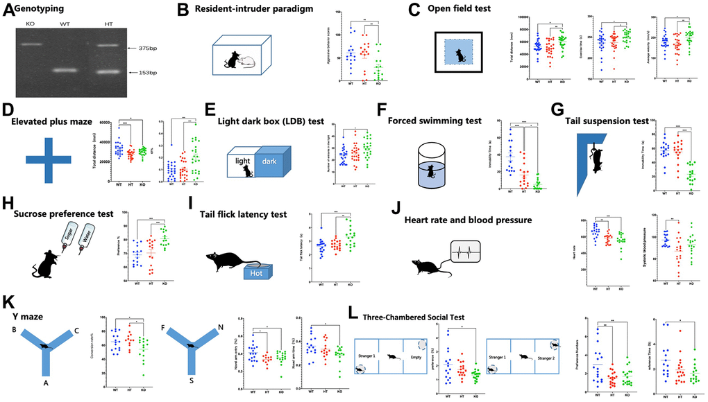
Figure 1. Genotyping, affective symptoms, and PMDD-like behaviors of mice. (A) Mouse genotyping using primers specific for the Gabrb2 and Neo genes. (B) Resident-intruder paradigm showing aggressive behavior scores of mice (WT male = 16, HT male= 16, KO male=16). (C) Open field test showing total distance, total distance in the central area, and total time in the central area (WT male =24, HT male=25, KO male=24). (D) Elevated plus maze showing percentile entries and time into open arms (WT male =24, HT male=25, KO male=24). (E) Light dark box (LDB) test showing entries and time into lightbox (WT male = 18, HT male= 18, KO male= 18). (F) Forced swimming test showing immobility time of mice suspended in the water (WT male =15, HT male=17, KO male=17). (G) Tail suspension test showing immobility time of mice suspended by the tail to a horizontal bar (WT male =17, HT male=17, KO male=17). (H) Sucrose preference test showing (WT male =15, HT male=16, KO male=15). (I) Tail flick latency test showing (WT male =18, HT male= 17, KO male=16). (J) Heart rate and blood pressure (WT male =18, HT male=18, KO male=18). (K) Y maze contains two parts: spontaneous alternation and novelty arm, showing percentile time spent in, or entries into, the novel arm was monitored to measure spatial-working memory (WT male =18, HT male=12, KO male= 17). (L) Three-Chambered Social Test contains two parts: social affiliation and social novelty, showing a preference for the container holding a stranger mouse relative to an empty container; and preference for social novelty: preference for the container holding a Stranger-2 mouse relative to the container holding Familiar-1 mouse (WT male =18, HT male=18, KO male= 18). Statistical analysis was performed using one-way ANOVA with Newman–Keuls post-hoc test. Average y values ± SEM in the different plots are represented by horizontal bars. *p < 0.05, **p < 0.01, ***p < 0.001.
Behavioral tests
Male mice (8-10 weeks) were selected for behavioral tests according to previous research [22, 23]. In the experiment, the “open field test-elevated plus maze-light and dark box” were used for multi-aspect evaluation of the anxiety-like emotional performance. The forced swimming-tail suspension-sucrose preference tests were adopted for multi-aspect assessment of depression-like dynamic performance. The Y-maze test was employed to examine mice's learning and memory capacities. The sociability of mice was assessed using the three-chamber sociability test. The resident intruder paradigm and the light-heat flick were used to evaluate mice’s irritable aggression behavior and pain sensitivity. Lastly, heart rate and blood pressure were measured to examine the activity of sympathetic nerves. Specific procedures are presented in the Supplementary Materials.
Measurement of GABAAR subunits protein levels in Gabrb2-knockout mice
Western blot assay was used to measure the expression patterns of GABAAR Subunits protein in brain tissues of mice. Radio-immunoprecipitation assay (RIPA) buffer for protein extraction. Brain tissues were comprehensively lysed and homogenized. The protein concentration was determined and modified to appropriate attention for protein expression detection. Subsequent procedures were performed as shown in the Supplementary Materials. The Image Lab 5.2.1 software was employed for analysis.
Neurotransmitter detection in Gabrb2-knockout mice
A HILIC-MS/MS method was adopted to detect neurotransmitter levels in serum as well as the brain tissues of mice according to the procedures described in the Supplementary Materials.
Electrophysiological recordings
After anesthesia, the abdominal cavities of the animals were opened, and the chest opened upward to expose the heart. Using a syringe, pre-cooled artificial cerebrospinal fluid (ACSF) was injected into the aorta via the left ventricle; the auricular appendix was after that incised for perfusion. After cervical dislocation, the skin was marked, and the skull was opened using a scalpel to expose the whole brain tissues. A medicine spoon was used to harvest the brain tissues and allowed to stand in pre-cooled ACSF (filled with mixed air to saturated condition) for 2 minutes. The brain tissues were placed in a petri dish paved with filter paper. Meanwhile, the ACSF and mixer were transferred onto the petri dish. The tissues containing the cortex were excised using a blade, then glued in the correct direction on the agar-coated plate of the brain-slicing machine. Subsequently, the tissues were immediately transferred to the slicing slot of the brain-slicing machine, fixed, then sectioned into slices of 250 μM thickness. For the cortical regions, specific hippocampal areas of the brain slices were cut off and digested with trypsin (formulated with HBSS) at 33.3° C for 30 min, under high purity oxygen exposure. The trypsin was removed after digestion; after that its effect was terminated by loading each tube with 3 mL oxygenated HBSS Na+ solution. This operation was repeated three times. Subsequently, 2 mL of oxygenated Low Ca2+ HEPES was added, then the tissues were triturated using a Pasteur pipet in the large, medium, and small diameters. After trituration by a Pasteur pipet in large, medium diameters, the tissues were allowed to stand for 2 min. The supernatant was pipetted into a new 15 mL centrifuge tube, followed by 4-5 cycles of trituration with a Pasteur pipet in a small diameter. The mixture was allowed to stand for 2 min then the supernatant was obtained and mixed with the former supernatant harvested above. The mixed cell suspension was seeded into a 3.5 cm polylysine-coated petri dish, 2 mL/dish. The cells were left to stand for 10 min to allow cell adherence to the wall.
Statistical analysis
All statistical analyses were performed using the IBM SPSS statistical 22 software. Data were expressed in mean ± standard deviation. Comparisons between two groups were performed using one sample unpaired t-test, whereas comparisons between multiple groups were conducted using a one-way analysis of variance (ANOVA). The GABAA receptor current in response to drug treatment and the control GABA assessment (20 μM) were adjusted for data obtained after an electrophysiological examination. Further, we calculated the enhancement drug ratio to the GABAA receptor current. The values of *p < 0.05, **p < 0.01, ***p < 0.001 were considered statistically significant. The GraphPad Prism 8.4 software was used for image plotting.
Data availability
The data supporting the findings of this study are accessible by the corresponding author upon request.
Results
Changes in the emotional state of Gabrb2 knock-out mice
Assessment of anger-like emotions revealed that the aggressive behavioral scores of Gabrb2 KO mice were significantly lower than that of WT mice (p < 0.01, Figure 1B). The anxiety-like emotions were assessed by open field test (OFT), elevated plus maze (EPM), and light-dark box, which revealed significantly longer total distance and increased mean velocity of Gabrb2 KO mice compared to the WT mice (p < 0.05) and HT mice (p < 0.01) and prolonged movement time as compared to the WT mice (p < 0.05) and HT mice (p < 0.05) in the OFT. Substantial shortened total movement distance was measured in KO mice versus HT mice (p < 0.001) and WT mice (p < 0.01), whereas notable elevated OT% was found in Gabrb2 KO mice versus WT mice (p < 0.001) and WT mice (p < 0.001). In the light-dark box, the entrance times into the bright area were markedly increased in Gabrb2 KO mice than that in WT mice (p < 0.01) but similar to that in HT mice (p > 0.05). The depression-like emotions were evaluated by the tail suspension test (TST), FST, and a sucrose preference test; consequently, the immobility time in TST significantly decreased in Gabrb2 KO mice compared with that in WT mice (p < 0.001) and HT mice (p < 0.001). The immobility time in the FST of Gabrb2 KO mice was shortened compared to that of WT mice (p < 0.001) and HT mice (p < 0.05), whereas that of HT mice was noticeably shorter than that of WT mice (p < 0.001). The sucrose preference index of Gabrb2 KO mice was higher than that of WT mice (p < 0.001) and HT mice (p < 0.001), (Figure 1C–1H).
Changes in the somatic state of Gabrb2 knock-out mice
Further, we evaluated the activity of sympathetic nerves and pain thresholds of mice. Consequently, the KO mice showed a lower heart rate than WT mice (p < 0.001), whereas HT mice had a lower heart rate than WT mice (p < 0.01). No difference in the diastolic blood pressure was observed among the groups. Nevertheless, the systolic blood pressure of HT mice was remarkably lower than that of WT mice (p < 0.01). The tail-flick test demonstrated that the pain threshold of KO mice was notably higher than that of WT mice (p < 0.01) and that of HT mice (p < 0.001) (Figure 1I, 1J).
GABAAR receptor pathway changes in the Gabrb2 knock-out mice
Western blot assay was used to measure the expression patterns of GABAA receptor α1-6 subunits (GABRA1, GABRA2, GABRA3, GABRA4, GABRA5, and GABRA6), β1-3 subunits (GABRB1, GABRB2, and GABRB3), γ1-3 subunits (GABRG1, GABRG2, and GABRG3), δ subunit (GABRD), ε subunit (GABRE), π subunit (GABRP), and θ subunit (GABRQ). Protein levels of GABAA receptors δ and ε subunits were substantially increased in the cerebral regions. GABAA receptor δ subunit protein expression significantly increased in the cerebellum of Gabrb2 KO mice compared to WT mice (p < 0.05), (Figure 2).
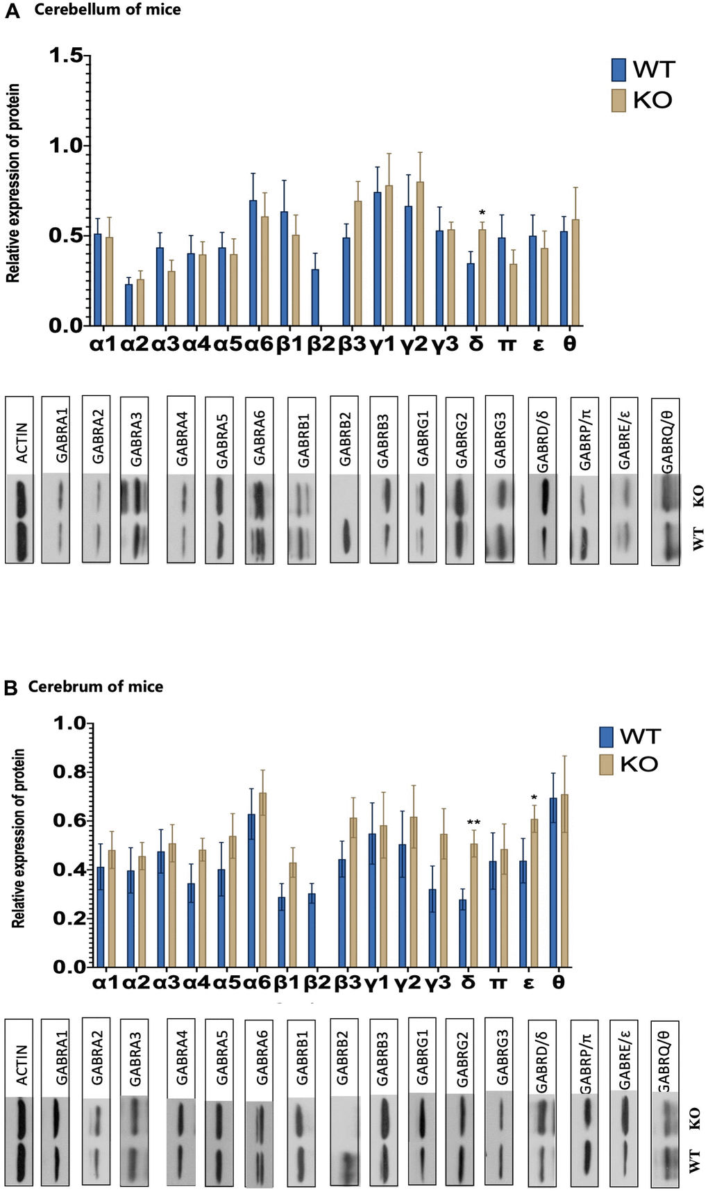
Figure 2. GABAAR receptor subunits changes. (A) The levels of protein expression for 16 different GABA A receptor subunits of WT and KO mouse in the cerebrum (WT male =8, KO male= 8). (B) The levels of protein expression for 16 different GABA A receptor subunits of WT and KO mouse in the cerebellum (WT male =8, KO male=8). Statistical analysis was performed using one-way ANOVA with Newman–Keuls post-hoc test. Average y values ± SEM in the different plots are represented by horizontal bars. *p < 0.05, **p < 0.01, ***p < 0.001.
Further, we evaluated the levels of 4-aminobutyric acid (GABA), glutamic acid, dopamine, serotonin, norepinephrine, and epinephrine in the peripheral blood of WT and Gabrb2 KO mice. As a result, the levels of 4-aminobutyric acid (GABA), glutamic acid, serotonin, and norepinephrine in the peripheral blood of KO mice were similar to that in WT mice (p > 0.05). Nonetheless, the levels of dopamine and epinephrine in the peripheral blood of Gabrb2 KO mice were strikingly higher than that of WT mice (p < 0.05). The contents of glutamic acid, norepinephrine, serotonin, and epinephrine revealed insignificant differences in the cerebrum tissues of Gabrb2 KO mice and WT mice (p > 0.05). The GABA, GABA/Glu, and dopamine levels were noticeably increased, whereas that of acetylcholine decreased in the cerebral tissues of Gabrb2 KO mice relative to WT mice (p < 0.05). The acetylcholine was not detected in the cerebellum of Gabrb2 KO mice, and the levels of dopamine and serotonin in the cerebellum of Gabrb2 KO mice were similar to that of WT mice (p > 0.05). The levels of 4-aminobutyric acid (GABA), glutamic acid, norepinephrine, and epinephrine were significantly higher in the Gabrb2 KO mice than that in the WT mice (p < 0.05). These data indicate that Gabrb2 KO causes compensatory changes in GABA, dopamine, and acetylcholine. Also, GABA/Glu in the cerebral regions may be related to the behavioral phenotype of transgenic mice (Figure 3).
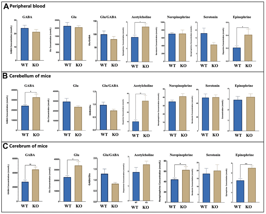
Figure 3. Neurotransmitters changes. (A) The level of key neurotransmitters in vivo of WT and KO mouse (WT male =8, KO male= 8). (B) The levels of WT and KO mouse in the cerebrum (WT male =8, KO male= 8). (C) The levels of neurotransmitters of WT and KO mouse in the cerebellum (WT male =8, KO male= 8). The levels of GABA, Glu Statistical analysis was performed using one-way ANOVA with Newman–Keuls post-hoc test. Average y values ± SEM in the different plots are represented by horizontal bars. *p < 0.05, **p < 0.01, ***p < 0.001.
Dose effect of ALLO on Gabrb2 KO mice
The behavioral and cell level changes were observed in mice injected with ALLO. The overall distance of EPM was shortened in a dose-dependent manner after ALLO injection into mice with three genotypes. In response to treatment with 20 mg/kg ALLO, the WT mice significantly covered a shortened total distance. Also, ALLO treatment at doses of 17 mg/kg and 20 mg/kg significantly shortened the total movement distance of HT mice (p < 0.01). ALLO treatment at a dose of 20 mg/kg markedly shortened the total movement distance of Gabrb2 KO mice (p < 0.01). Additionally, ALLO treatment at a dose of 17 mg/kg considerably increased the OT% of WT mice and HT mice (p < 0.01), and a dose of 20 mg/kg noticeably increased the OT% of KO mice (p < 0.01). Furthermore, 17 mg/kg ALLO augmented the OE% of WT mice (p < 0.05), whereas 10 mg/kg and 20 mg/kg ALLO significantly augmented the OE% of WT mice (p < 0.01). In the TST test, the mice of three genotypes were injected with different doses of ALLO. The immobility time of HT mice was unaffected (p > 0.05), whereas that of WT mice was prolonged following treatment with different doses of ALLO, among which 20 mg/kg had a significant effect (p < 0.01). ALLO treatment at different doses prolonged the immobility time of Gabrb2 KO mice; unlike 10 mg/kg dose (p > 0.05), 17 mg/kg and 20 mg/kg doses exerted a significant effect (p < 0.01), (Figure 4A, 4B).
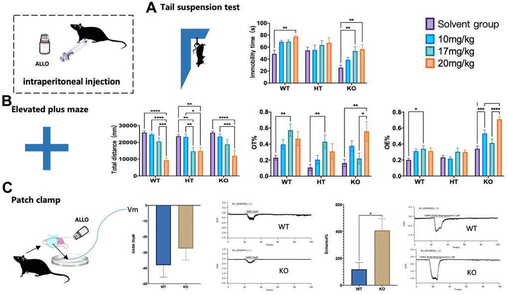
Figure 4. Changes of KO behavioral phenotypes by ALLO. The behaviors of WT, HT, or KO mice administered with 10 mg/kg, 17 mg/kg, 20 mg/kg ALLO i.p was compared with that of control mice administered with saline. (A) Tail suspension test showing immobility time of mice suspended by the tail to a horizontal bar (WT male: saline=12, 10 mg/kg=12, 17 mg/kg=12, 20 mg/kg=12; HT male: saline= 12,10 mg/kg=11, 17 mg/kg=12, 20 mg/kg=12; KO male: saline=11,10 mg/kg=12, 17 mg/kg=11 20 mg/kg=10). (B) Elevated plus maze showing percentile entries and time into open arms (WT male: saline=12, 10 mg/kg=12, 17 mg/kg=12, 20 mg/kg=10; HT male: saline= 12,10 mg/kg=11, 17 mg/kg=12, 20 mg/kg=11; KO male: saline=12,10 mg/kg=12, 17 mg/kg=12, 20 mg/kg=12). (C) Patch clamp showing changes of GABAA receptor current in mouse cortical neurons under ALLO intervention between WT male mice and KO male mice (WT male =6, HT male=6, KO male=6). Statistical analysis was performed using one-way ANOVA with Newman–Keuls post-hoc test. Average y values ± SEM in the different plots is represented by horizontal bars. *p < 0.05, **p < 0.01, ***p < 0.001.
The patch-clamp technique was used to assess the allosteric regulation of ALLO on GABAA receptor currents in the cortical neurons of WT and Gabrb2 KO mice. No difference in receptor currents was noted in the mouse cerebral cortex in response to treatment with 20 μM GABA. Nevertheless, 1 μM ALLO induced a more significant change in cerebral cortex current in Gabrb2 KO mice than that in WT mice. ALLO enhanced the agonistic effect of the GABAARs in cortical neurons in the context of β2 subunit deletion. Therefore, GABAARβ2 subunit deletion did not affect the binding to ALLO. We could not ascertain whether the ALLO binding site was located in the β2 subunit. However, abnormal expression of the GABAARβ2 subunit certainly caused changes in other subunits, thereby affecting the binding to ALLO. This results in abnormal changes in cerebral receptor currents, manifesting as abnormal behavioral changes in mice (Figure 4C).
Discussion
According to the published relevant research, the open-field test (OFT) is used to test for behavioral symptoms of PMDD rat models; elevated plus maze (EPM) and light dark box (LDB) tests for anxiety [24]; resident-intruder paradigm for irritability [25]; forced swimming test for depression [26]; saccharin preference test for anhedonia; social preference-avoidance test for social withdrawal [27], heart rate (HR) and blood pressure (BP) [28]. As expected, Gabrb2 KO mice displayed changes in anxiety-like and depression-like emotions in contrast with PMDD symptoms; changes in the social, learning, and memory abilities similar to PMDD symptoms; changes in pain threshold opposite to PMDD symptoms, hence reducing pain sensitivity. The results above corroborate with the previous studies, which focused on schizophrenia-like phenotypes [22]. Our study concentrated on PMDD-like phenotypes and added new tests for assessing pain sensitivity and heart rate/blood pressure related to PMDD [28–30]. These findings confirm that protein function mediated by Gabrb2 is closely associated with the pathogenesis of PMDD.
Meanwhile, we adopted molecular biotechnology to analyze the compensatory changes of Gabrb2 KO mice. Recent research shows that anxiety may result in abnormal expression of α4β2δ GABAAR [31]. Among GABAAR subunits, GABAAR δ and GABAAR ε subunit proteins were expressed at higher abundance in the brain region of the Gabrb2 KO mice. In contrast, GABAA receptor δ subunit proteins were significantly expressed in the cerebellar areas. Previous research indicates that corticotropin-releasing hormone (CRH) neurons are modulated by neurosteroid tetrahydro-deoxycorticosterone (THDOC) and act on GABAAR-containing δ subunits which share close associations with anxiety-like behaviors [32]. The δ-Subunit is necessary for Protein Kinase C-dependent effects of neurosteroids on synaptic GABA A receptor inhibition [33]. Besides, GABAA(δ)R may promote fear extinction via a route relying on non-synaptic plasticity [34]. Therefore, GABAA(δ)R has high research potential. Moreover, we found a compensatory increase in dopamine and epinephrine levels in the peripheral blood of transgenic mice; compensatory changes in GABA, dopamine, acetylcholine, and GABA/Glu levels in the brain regions; compensatory changes in GABA, glutamic acid, norepinephrine, epinephrine, and GABA/Glu in the cerebellar area of the mice. Therefore, we speculate that most of the above-stated changes may correlate with the behavioral phenotype of transgenic mice. Besides GABA and Glu indicated above, 5-HT, a monoamine neurotransmitter regulates emotions and cognitive functions in the CNS [35]. NE, DA, etc., are important indicators for assessing depression severity [36]. Also, research has demonstrated a strong correlation between dopamine, 5-HT, norepinephrine, and serotonin in the brain with depression-like emotions [37], which is in agreement with our findings [38].
The neuroactive steroid allopregnanolone (ALLO) is an endogenous positive allosteric modulator of GABA type A receptor (GABAAR), which causes the development of mood disorders, including depression, anxiety, and PMDD [39–41]. Nonetheless, it remains unclear whether ALLO mediates Gabrb2 as a significant subunit [42]. When ALLO was injected into animal experiments, Gabrb2 KO did not yield the expected effect on the mouse phenotype. Unlike in the WT mice, ALLO treatment improved the agonistic effect on the GABAA receptor in cortical neurons of KO mice, supporting the hypothesis that the ALLO binding site may not be located on the β2 subunit. Changes in mice caused by abnormal expression of β2 subunit may be related to ALLO sensitivity. However, there is an urgent need for additional comprehensive studies.
Conclusions
GABAAR β 2 has a regulatory effect on PMDD-like behaviors with disturbance of neurotransmitters, hence directly or indirectly affecting its mediated ALLO binding in vital brain areas. This causes abnormal nerve synaptic currents, and manifestation of PMDD-related clinical symptoms, which should be validated in future research.
Author Contributions
Mingzhou Gao designed experiments. Mingzhou Gao, Hao Zhang, Ya Sun, Zhan Gao and Sunyan Sun performed animal experimental modeling and detection of biological indexes. Fengqin Wei and Dongmei Gao provided the funding and key assistance. Mingzhou Gao performed statistics and completed the manuscript with Hao Zhang. All authors read and approved the manuscript.
Acknowledgments
We are grateful to Xue Hong’s guidance from the Hong Kong University of science and technology, and the experimental platform and experimental conditions provided by the Research and Innovation team of Emotional Diseases and Syndromes in Shandong University of Traditional Chinese Medicine led by Mingqi Qiao and all team members for their help in the experiment.
Conflicts of Interest
The authors declare that they have no conflicts of interest.
Ethical Statement and Consent
All animal protocols and procedures were approved by the Ethics committee of Shandong University of Traditional Chinese Medicine (SDUTCM20190904013) and were performed in accordance with the Institute for Laboratory Animal Research Guide for the Care and Use of Laboratory Animals.
Funding
This study was sponsored by Key Project of Natural Science Foundation of Shandong Province (ZR2020ZD17), National Natural Science Foundation of China (82204958), Traditional Chinese medicine science and technology project of Shandong Province (Q-2022059) and Medical and health science and technology development plan project of Shandong Province (202105010467).
References
- 1. Mishra S, Elliott H, Marwaha R. Premenstrual Dysphoric Disorder. StatPearls. Treasure Island (FL): StatPearls Publishing. 2022. [PubMed]
- 2. Gao M, Gao D, Sun H, Cheng X, An L, Qiao M. Trends in Research Related to Premenstrual Syndrome and Premenstrual Dysphoric Disorder From 1945 to 2018: A Bibliometric Analysis. Front Public Health. 2021; 9:596128. https://doi.org/10.3389/fpubh.2021.596128 [PubMed]
- 3. Tiranini L, Nappi RE. Recent advances in understanding/management of premenstrual dysphoric disorder/premenstrual syndrome. Fac Rev. 2022; 11:11. https://doi.org/10.12703/r/11-11 [PubMed]
- 4. Delara M, Borzuei H, Montazeri A. Premenstrual disorders: prevalence and associated factors in a sample of Iranian adolescents. Iran Red Crescent Med J. 2013; 15:695–700. https://doi.org/10.5812/ircmj.2084 [PubMed]
- 5. Daşıkan Z. Premenstrual disorders among young Turkish women: According to DSM-IV and DSM-V criteria using the premenstrual symptoms screening tool. Perspect Psychiatr Care. 2021; 57:481–7. https://doi.org/10.1111/ppc.12656 [PubMed]
- 6. Perkonigg A, Yonkers KA, Pfister H, Lieb R, Wittchen HU. Risk factors for premenstrual dysphoric disorder in a community sample of young women: the role of traumatic events and posttraumatic stress disorder. J Clin Psychiatry. 2004; 65:1314–22. https://doi.org/10.4088/jcp.v65n1004 [PubMed]
- 7. O’Brien S. Premenstrual syndrome/premenstrual dysphoric disorder: Pathophysiology, definition of the disease and treatment options. Journal of Psychosomatic Obstetrics and Gynecology. 2007; 28:55.
- 8. Shams-Alizadeh N, Maroufi A, Rashidi M, Roshani D, Farhadifar F, Khazaie H. Premenstrual dysphoric disorder and suicide attempts as a correlation among women in reproductive age. Asian J Psychiatr. 2018; 31:63–6. https://doi.org/10.1016/j.ajp.2018.01.003 [PubMed]
- 9. Osborn E, Brooks J, O’Brien PM, Wittkowski A. Suicidality in women with Premenstrual Dysphoric Disorder: a systematic literature review. Arch Womens Ment Health. 2021; 24:173–84. https://doi.org/10.1007/s00737-020-01054-8 [PubMed]
- 10. Hantsoo L, Epperson CN. Allopregnanolone in premenstrual dysphoric disorder (PMDD): Evidence for dysregulated sensitivity to GABA-A receptor modulating neuroactive steroids across the menstrual cycle. Neurobiol Stress. 2020; 12:100213. https://doi.org/10.1016/j.ynstr.2020.100213 [PubMed]
- 11. Bixo M, Johansson M, Timby E, Michalski L, Bäckström T. Effects of GABA active steroids in the female brain with a focus on the premenstrual dysphoric disorder. J Neuroendocrinol. 2018; 30. https://doi.org/10.1111/jne.12553 [PubMed]
- 12. Epperson CN. Premenstrual dysphoric disorder and the brain. Am J Psychiatry. 2013; 170:248–52. https://doi.org/10.1176/appi.ajp.2012.12121555 [PubMed]
- 13. Syan SK, Minuzzi L, Smith M, Costescu D, Allega OR, Hall GB, Frey BN. Brain Structure and Function in Women with Comorbid Bipolar and Premenstrual Dysphoric Disorder. Front Psychiatry. 2018; 8:301. https://doi.org/10.3389/fpsyt.2017.00301 [PubMed]
- 14. Kaltsouni E, Fisher PM, Dubol M, Hustad S, Lanzenberger R, Frokjaer VG, Wikström J, Comasco E, Sundström-Poromaa I. Brain reactivity during aggressive response in women with premenstrual dysphoric disorder treated with a selective progesterone receptor modulator. Neuropsychopharmacology. 2021; 46:1460–7. https://doi.org/10.1038/s41386-021-01010-9 [PubMed]
- 15. Smith SS, Shen H, Gong QH, Zhou X. Neurosteroid regulation of GABA(A) receptors: Focus on the alpha4 and delta subunits. Pharmacol Ther. 2007; 116:58–76. https://doi.org/10.1016/j.pharmthera.2007.03.008 [PubMed]
- 16. Wang M. Neurosteroids and GABA-A Receptor Function. Front Endocrinol (Lausanne). 2011; 2:44. https://doi.org/10.3389/fendo.2011.00044 [PubMed]
- 17. Ullah A, Long X, Mat WK, Hu T, Khan MI, Hui L, Zhang X, Sun P, Gao M, Wang J, Wang H, Li X, Sun W, et al. Highly Recurrent Copy Number Variations in GABRB2 Associated With Schizophrenia and Premenstrual Dysphoric Disorder. Front Psychiatry. 2020; 11:572. https://doi.org/10.3389/fpsyt.2020.00572 [PubMed]
- 18. Huiyun Z, Jieqiong W, Heng L. Hippocampus of rat model of liver qi inversion in premenstrual syndrome γ- Aminobutyric acid A receptor β_ 2 subunit mRNA expression. Journal of Beijing University of Traditional Chinese Medicine. 2008; 608–10.
- 19. Barki M, Xue H. GABRB2, a key player in neuropsychiatric disorders and beyond. Gene. 2022; 809:146021. https://doi.org/10.1016/j.gene.2021.146021 [PubMed]
- 20. Zhang T, Li J, Yu H, Shi Y, Li Z, Wang L, Wang Z, Lu T, Wang L, Yue W, Zhang D. Meta-analysis of GABRB2 polymorphisms and the risk of schizophrenia combined with GWAS data of the Han Chinese population and psychiatric genomics consortium. PLoS One. 2018; 13:e0198690. https://doi.org/10.1371/journal.pone.0198690 [PubMed]
- 21. Hirose S. Mutant GABA(A) receptor subunits in genetic (idiopathic) epilepsy. Prog Brain Res. 2014; 213:55–85. https://doi.org/10.1016/B978-0-444-63326-2.00003-X [PubMed]
- 22. Yeung RK, Xiang ZH, Tsang SY, Li R, Ho TY, Li Q, Hui CK, Sham PC, Qiao MQ, Xue H. Gabrb2-knockout mice displayed schizophrenia-like and comorbid phenotypes with interneuron-astrocyte-microglia dysregulation. Transl Psychiatry. 2018; 8:128. https://doi.org/10.1038/s41398-018-0176-9 [PubMed]
- 23. Lovick TA, Zangrossi H
Jr . Effect of Estrous Cycle on Behavior of Females in Rodent Tests of Anxiety. Front Psychiatry. 2021; 12:711065. https://doi.org/10.3389/fpsyt.2021.711065 [PubMed] - 24. Zhang H, Geng X, Li Z, Li Y, Xu K, Wu H, Xie J, Sun P, Wei S, Qiao M. Paeonol at Certain Doses Alleviates Aggressive and Anxiety-Like Behaviours in Two Premenstrual Dysphoric Disorder Rat Models. Front Psychiatry. 2020; 11:295. https://doi.org/10.3389/fpsyt.2020.00295 [PubMed]
- 25. Wei S, Sun P, Guo Y, Chen J, Wang J, Song C, Li Z, Xue L, Qiao M. Gene Expression in the Hippocampus in a Rat Model of Premenstrual Dysphoric Disorder After Treatment With Baixiangdan Capsules. Front Psychol. 2018; 9:2065. https://doi.org/10.3389/fpsyg.2018.02065 [PubMed]
- 26. Wei S, Geng X, Li Z, Xu K, Hu M, Wu H, Shi W, Qiao M. A forced swim-based rat model of premenstrual depression: effects of hormonal changes and drug intervention. Aging (Albany NY). 2020; 12:24357–70. https://doi.org/10.18632/aging.202249 [PubMed]
- 27. Li Y, Pehrson AL, Budac DP, Sánchez C, Gulinello M. A rodent model of premenstrual dysphoria: progesterone withdrawal induces depression-like behavior that is differentially sensitive to classes of antidepressants. Behav Brain Res. 2012; 234:238–47. https://doi.org/10.1016/j.bbr.2012.06.034 [PubMed]
- 28. Girdler SS, Leserman J, Bunevicius R, Klatzkin R, Pedersen CA, Light KC. Persistent alterations in biological profiles in women with abuse histories: influence of premenstrual dysphoric disorder. Health Psychol. 2007; 26:201–13. https://doi.org/10.1037/0278-6133.26.2.201 [PubMed]
- 29. Landén M, Wennerblom B, Tygesen H, Modigh K, Sörvik K, Ysander C, Ekman A, Nissbrandt H, Olsson M, Eriksson E. Heart rate variability in premenstrual dysphoric disorder. Psychoneuroendocrinology. 2004; 29:733–40. https://doi.org/10.1016/S0306-4530(03)00117-3 [PubMed]
- 30. Poromaa IS. Physiological Correlates of Premenstrual Dysphoric Disorder (PMDD). Curr Top Behav Neurosci. 2014; 21:229–43. https://doi.org/10.1007/7854_2014_296 [PubMed]
- 31. Gallo AT, Hulse GK. A theory of the anxiolytic action of flumazenil in anxiety disorders. J Psychopharmacol. 2022; 36:439–48. https://doi.org/10.1177/02698811221082466 [PubMed]
- 32. Sarkar J, Wakefield S, MacKenzie G, Moss SJ, Maguire J. Neurosteroidogenesis is required for the physiological response to stress: role of neurosteroid-sensitive GABAA receptors. J Neurosci. 2011; 31:18198–210. https://doi.org/10.1523/JNEUROSCI.2560-11.2011 [PubMed]
- 33. Littlejohn EL, Boychuk CR. Protein Kinase C-Dependent Effects of Neurosteroids on Synaptic GABAA Receptor Inhibition Require the δ-Subunit. Front Physiol. 2021; 12:742838. https://doi.org/10.3389/fphys.2021.742838 [PubMed]
- 34. Pan HQ, Liu XX, He Y, Zhou J, Liao CZ, You WJ, Jiang SY, Qin X, Chen WB, Fei EK, Zhang WH, Pan BX. Prefrontal GABAA(δ)R Promotes Fear Extinction through Enabling the Plastic Regulation of Neuronal Intrinsic Excitability. J Neurosci. 2022; 42:5755–70. https://doi.org/10.1523/JNEUROSCI.0689-22.2022 [PubMed]
- 35. Okaty BW, Commons KG, Dymecki SM. Embracing diversity in the 5-HT neuronal system. Nat Rev Neurosci. 2019; 20:397–424. https://doi.org/10.1038/s41583-019-0151-3 [PubMed]
- 36. Meng P, Li C, Duan S, Ji S, Xu Y, Mao Y, Wang H, Tian J. Epigenetic Mechanism of 5-HT/NE/DA Triple Reuptake Inhibitor on Adult Depression Susceptibility in Early Stress Mice. Front Pharmacol. 2022; 13:848251. https://doi.org/10.3389/fphar.2022.848251 [PubMed]
- 37. Nutt DJ. Relationship of neurotransmitters to the symptoms of major depressive disorder. J Clin Psychiatry. 2008; 69:4–7. [PubMed]
- 38. Blier P. Neurotransmitter targeting in the treatment of depression. J Clin Psychiatry. 2013 (Suppl 2); 74:19–24. https://doi.org/10.4088/JCP.12084su1c.04 [PubMed]
- 39. Chen S, Gao L, Li X, Ye Y. Allopregnanolone in mood disorders: Mechanism and therapeutic development. Pharmacol Res. 2021; 169:105682. https://doi.org/10.1016/j.phrs.2021.105682 [PubMed]
- 40. Schiller CE, Schmidt PJ, Rubinow DR. Allopregnanolone as a mediator of affective switching in reproductive mood disorders. Psychopharmacology (Berl). 2014; 231:3557–67. https://doi.org/10.1007/s00213-014-3599-x [PubMed]
- 41. Bäckström T, Bixo M, Johansson M, Nyberg S, Ossewaarde L, Ragagnin G, Savic I, Strömberg J, Timby E, van Broekhoven F, van Wingen G. Allopregnanolone and mood disorders. Prog Neurobiol. 2014; 113:88–94. https://doi.org/10.1016/j.pneurobio.2013.07.005 [PubMed]
- 42. Bracamontes JR, Li P, Akk G, Steinbach JH. A neurosteroid potentiation site can be moved among GABAA receptor subunits. J Physiol. 2012; 590:5739–47. https://doi.org/10.1113/jphysiol.2012.237255 [PubMed]