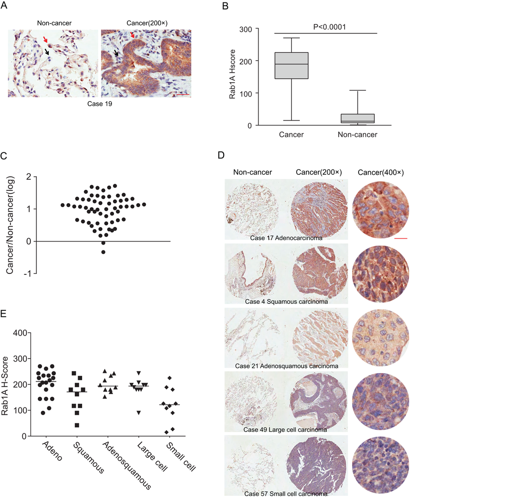
Figure 1.Rab1A is frequently overexpressed in different histological types of lung cancer. (A) IHC staining of human lung cancer tissue and noncancerous tissues. Shown are representative images of stained tumor and non-cancerous tissue sections. Red arrowhead: high Rab1A staining; black arrowhead: low Rab1A staining. Scale bar = 50 µm. (B) Box plot graph showing the statistical analysis of Rab1A expression in lung cancer and paired non-cancerous tissues. (C) Scatter plot showing Rab1A staining levels in individual tumors as a ratio of Rab1A staining in lung cancer to the paired non-cancerous tissue. (D) IHC staining of human lung cancer tissue microarray and paired noncancerous tissues. Shown are stained tumor and non-cancerous tissue sections representative of different histological types. Scale bar = 20 µm. (E) Scatter plot showing levels of Rab1A staining in different histological types of lung cancer as a ratio of Rab1A staining in cancer tissues to paired non-cancerous tissue.