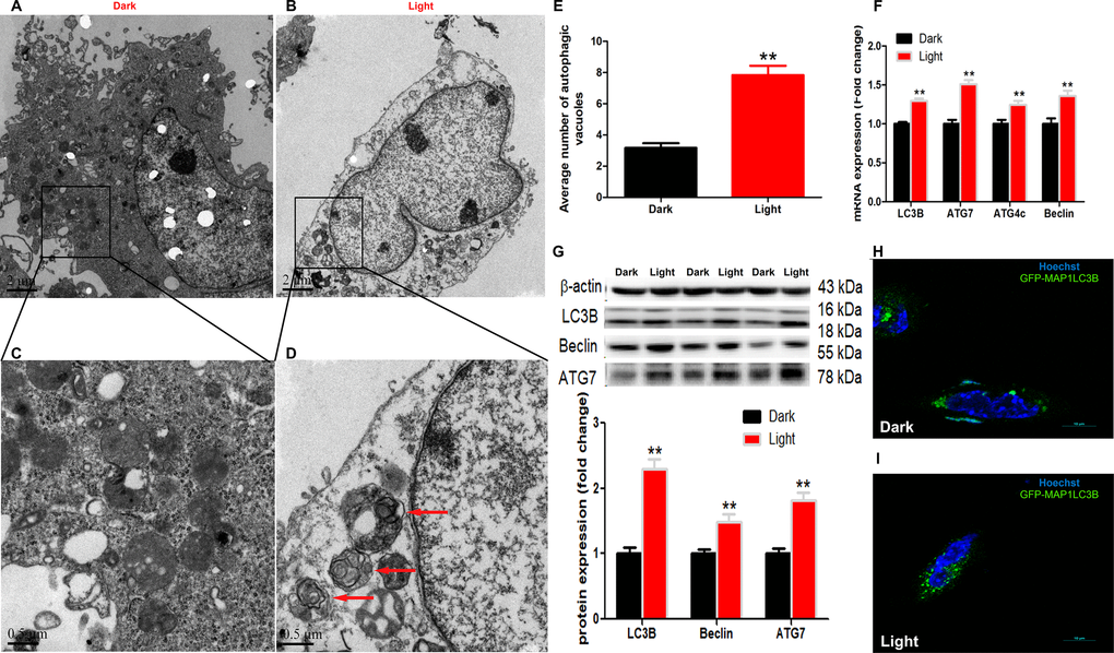
Figure 1.White LED light induces activation of autophagy process in HT-22 cells. (A–B) TEM images of autophagic vacuoles in Dark and Light groups, respectively. Scale bars, 2 μm, n = 3; (C–D) Higher magnification of indicated regions in A and B (squares), showing autophagosomes with double membrane (red arrows). Scale bars, 0.5 μm; (E) The number of autophagic vacuoles. Values are means ± SEM, ** p < 0.01 compared with Dark group, n = 3. (F) Quantitative Real-time PCR analysis of autophagy-related gene lc3b, atg7, atg4c and beclin. Values are means ± SEM, ** p < 0.01 compared with Dark group, n = 6; (G) Western blot analysis of autophagy-related protein LC3B, Beclin and ATG7. Values are means ± SEM, ** p < 0.01 compared with Dark group, n = 6; (H–I) Representative fluorescence images of HT-22 cells transfected with GFP-LC3 plasmid. Cells were counterstained with Hochest (nuclei in blue) and more autophagosomes (green) were seen in Light group. Scale bar, 10 μm.