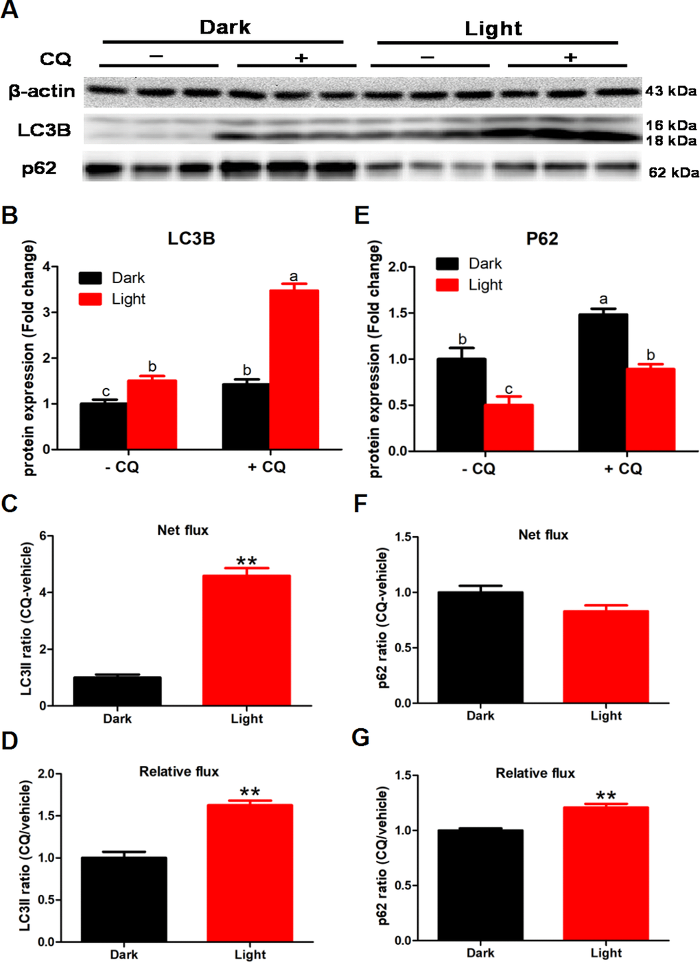
Figure 2.White LED light exposure enhance autophagy flux in hippocampal neuron cells. To detect the effect of white LED light on autophagy flux, we added 50 μM chloroquine (CQ) in the last 2 h of white LED light exposure. (A) Images of bands detected in Western blot analyses; (B) LC3B protein levels in Dark and Light group with or without CQ. Values are means ± SEM. Bars with different superscripts are significantly different from each other (p < 0.05, n = 3); (C) Net flux of LC3II protein. Values are means ± SEM, ** p < 0.01 compared with Dark group, n = 3; (D) Relative flux of LC3II protein. Values are means ± SEM, ** p < 0.01 compared with Dark group, n = 3; (E) p62 protein levels in Dark and Light group with or without CQ. Values are means ± SEM. Bars with different superscripts are significantly different from each other (p < 0.05, n = 3); (F) Net flux of p62 protein. Values are means ± SEM, n = 3; (G) Relative flux of p62 protein. Values are means ± SEM, ** p < 0.01 compared with Dark group, n = 3.