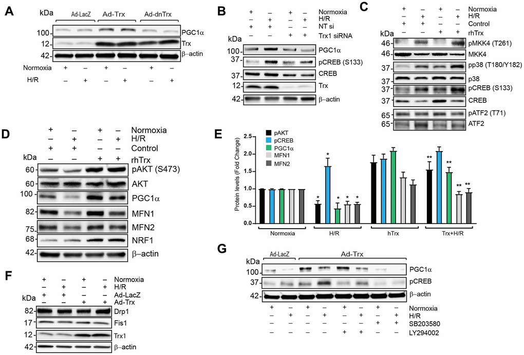
Figure 7.Trx regulates expression of PGC1α via PI3K-AKT-CREB axis in cardiomyocytes. (A). Western blot analysis of PGC1α, Trx and β-actin in lysate from Ad-LacZ, Ad-Trx and Ad-dnTrx infected and H/R (24/2h) exposed HCAECs. (B). Western blot analysis of PGC1α, pCREB (S133), CREB, Trx and b-actin in lysates from NT or Trx siRNA transfected and H/R (24/2h) exposed HCAECs. (C and D). Western blot analysis of pMKK4 (T261), MKK4, pp38 (T180/Y182), p38, pCREB (S133), CREB, pATF2 (T71), ATF2, pAKT (S473), AKT, PGC1α, MFN1, MFN2, NRF1 and β-actin in lysates from rhTrx pretreated and H/R (24/2h) exposed H9C2 cells. (E). Levels of protein in H9C2 cells were quantified and expressed as fold change. *p <0.05 versus Normoxia; **p <0.05 versus H/R. (F). Western blot analysis of Drp1, Fis1, Trx and β-actin in lysate from Ad-LacZ and Ad-Trx infected and H/R (24/2h) exposed H9C2 cells. (G). Western blot analysis of PGC1a, pCREB (S133) and β-actin in lysates from p38 inhibitor, SB203580 (2.5 μM) and PI3 kinase inhibitor, LY294002 (5.0 μM) pretreated and H/R (24/2h) exposed H9C2 cells. Statistical significance was determined with the Student’s t test.